3D Additive Manufacturing of Soft Materials as In Vitro Tumor Models
Participating Researchers
Current members: Dr. Meng Zhang, Moonkwang Jeong, Ann-Sophia Müller, Anita Stoppel, Anha Sara Maria Thaipadath Johnson
Former members: Nurcemal Atmaca, Dandan Li
Additive manufacturing enables the design and fabrication of objects with complex three-dimensional (3D) shapes that cannot be obtained from standard subtractive machining approaches, such as milling and etching. Additive technologies can therefore potentially revolutionize many fields, including biomedical technology. However, fast additive manufacturing schemes based on lithographic techniques are restricted to special materials, such as photopolymers. While optical techniques offer high resolution, it is often impossible to turn the built structures (e.g. photopolymers) into the desired material, especially if one requires structures that are soft and water-based. An alternative to lithography is direct 3D printing or jetting, but this results in a limited choice of materials and a limited resolution. Furthermore, direct printing methods are slow as they require the serial point by point writing of the structure.
The research project aims to develop the process to 3D print a soft printing material that simultaneously has high spatial resolution and high resemblance of real biological tissues. We demonstrate the range of potential applications by fabricating in vitro organ models with a resolution of tens of micrometers, including hollow branched networks that represent blood vessels, using materials, such as gels, that are soft and biocompatible [ref 1]. The method can generate realistic 3D complex environment with cells and extracellular matrices, which can be widely applied for in vitro experiments to test new biomedical devices and technologies.
Additive manufacturing of realistic organ models
Key collaborators: Prof. Arkadiusz Miernik, Department of Urology, Medical Center Freiburg
We built soft organ models by combining a commercial 3D printing technology with molding (Fig. 1). The detailed anatomical structure was first acquired from high resolution CT (computer tomography) data sets of human kidneys. CT reconstruction, ultrasound examination, and endoscopy showed that the soft phantom mimics a real kidney's detailed anatomy of both the outer shape and the collecting system with an average of 0.5 mm accuracy [ref2]. This method provides a cost-effective and precise model of the human organ in a reproducible and robust way. Moreover, it allows the usage of almost any kind of polymer materials that may mimic biological tissues including tumors [ref3], such as silicone materials (e.g. PDMS, Ecoflex) and hydrogels (e.g. agarose gel, gelatin) that can be used with real surgical instruments for realistic surgical simulation.
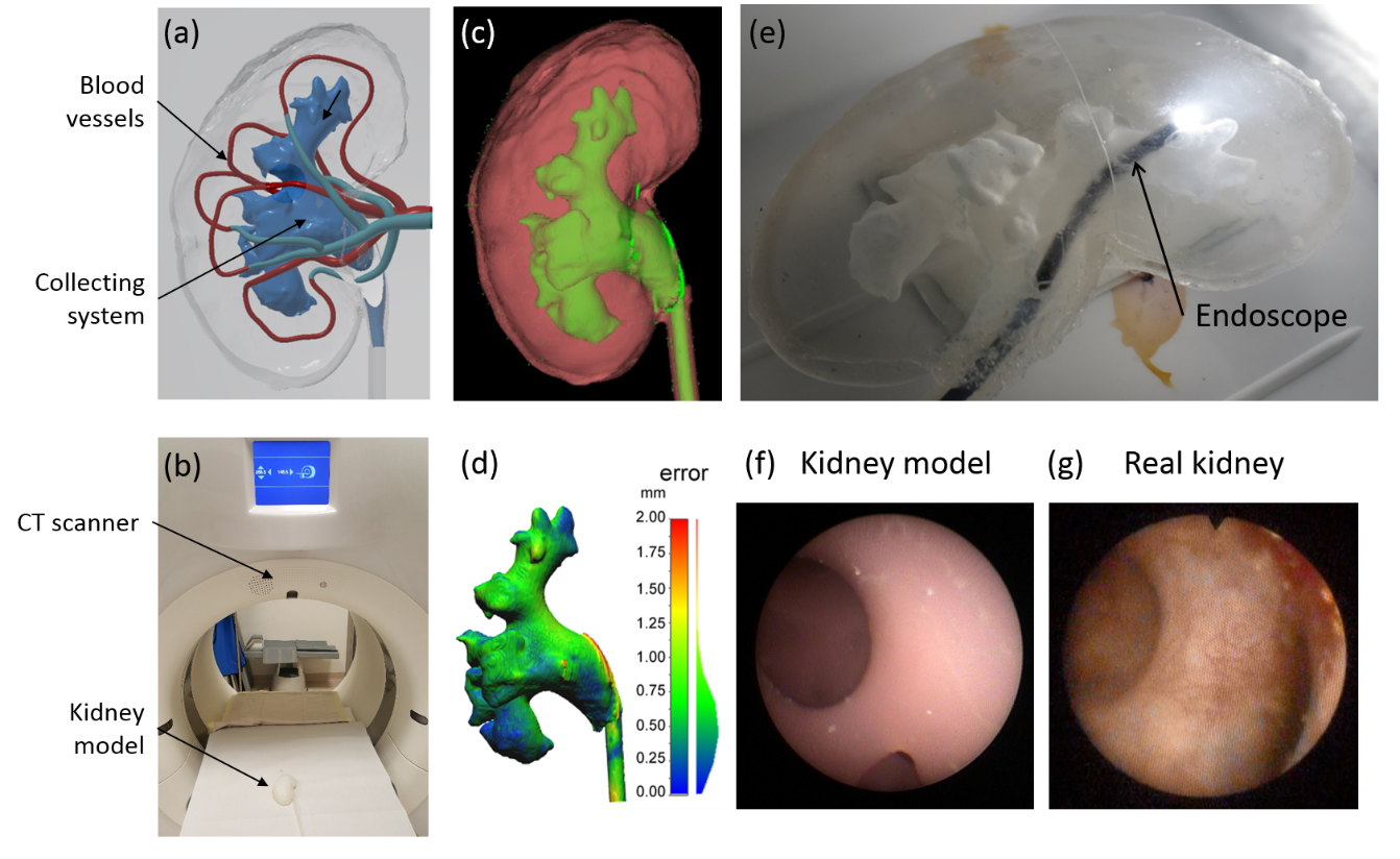
Fig 1. Validation of a human kidney model built using an additive manufacturing technique. (a) 3D model reconstructed based on medical CT data. (b) Validation of the kidney model with a CT scanner. (c) The reconstructed kidney model (the collecting system labeled in green and the surrounding tissue labeled in red). (d) Resolution analysis of the collecting system. (e) An endoscopic surgical procedure is simulated on the model. (f) and (g) The endoscopic view of the inside of the kidney model comparing with a real human kidney. [ref2]
© dkfz.de
Dynamic acoustic hologram for cell patterning
Key collaborators: Prof. Zhichao Ma, Shanghai Jiaotong University, China
Ultrasound actuation has mainly been shaped in resonator geometries, where the pressure patterns are a function of the resonator geometry and are thus highly symmetric. Alternately, several transducers are combined, and each transducer is controlled to have a defined phase. This can be extended to an array of several hundreds to a thousand transducers, known as a phased array, such that all the waves emanating from the transducers interfere to generate a defined pressure pattern. Phased arrays require complex instrumentation, as each transducer must be individually addressed.
We developed a radically new approach that enables the generation of complex 3D ultrasound fields with much higher complexity. This work has been published in Nature [ref4]. We used a 3D-printed highly-structured phase plate the acoustic hologram to achieve arbitrary shaped acoustic pressure fields. A plane wave coming from a single transducer that passes through the acoustic hologram element experiences a phase modulation across its wavefront. The 3D-printed acoustic hologram can be finely structured and can thus give rise to diffraction-limited arbitrary shaped acoustic pressure fields with unparalleled complexity. Recently, we also develop a new MEMS device for the dynamic modulation of an acoustic hologram, and demonstrated the capability of using ultrasound field to wirelessly manipulate soft microparticles and pattern cells (Fig. 2) [ref5].
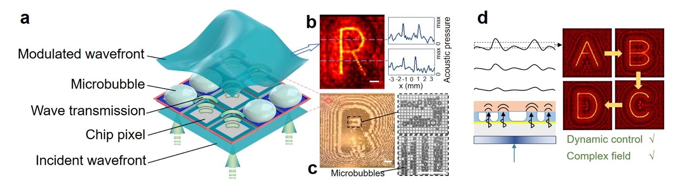
Figure 2. Spatial ultrasound modulator (SUM). (a) Schematics. A CMOS chip is used to form digitally controlled microbubble arrays. A bubble in water can block ultrasound, and it causes amplitude modulation of the wave. (b-c) Experimental pressure scan of the acoustic field in the target plane, controlled by the SUM. (d) An ultrasound movie is projected by refreshing the CMOS chip. [ref5]
© dkfz.de
Key publications
1) Atmaca N., et al. 3D Fabrication of Transparent Brain Phantom with Vascular Structure, in preparation.
2) Adams F., et al. (2016): ABME, kidney phantom.
3) Choi et al. (2021): Soft urinary bladder phantom for endoscopic training, Ann. Biomed. Eng., 49, 2412-2420
4) Melde, K. et al. (2016): Holograms for acoustics, Nature, 537, 518
5) Ma, Z. et al. (2020): Spatial ultrasound modulation by digitally controlling microbubble arrays, Nature Communications, 11, 4537