Ultrasound (sonography, abbr. US) a non-invasive examination method using sound waves
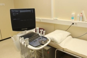
Ultrasound unit Zonare ZS3
© dkfz.de
Diagnostic ultrasound, a non-invasive and clinically extremely valuable examination using ultrasound waves, is a cross-section imaging modality like computed tomography. In real time, it produces live images of abdominal organs like spleen, liver, pancreas, or kidneys, but also of superficial organs like thyroid, parathyroid glands, head and neck soft tissue, breast, testicles, or lymph nodes without any hazard or discomfort to the patient. Since no x-rays are being used, ultrasound can be used for the pregnant as well as for neonates, infants, or children. Therefore, if you believe you could be pregnant, there will be no concern using ultrasound.
Ultrasound examinations at the DKFZ information on the procedure and valuable hints for the patient
Various ultrasound examinations are being performed at the DKFZ. Since air-filled bowel loops may obstruct the penetration of ultrasound waves into deeper-lying organs in the abdomen (like e.g. the pancreas), no heavy or bloating meals should be taken prior to the examination. For certain examinations, you should even be fasting. Please contact your referring doctor or our reception staff.
Depending on the organ to be examined, the skin above it should be exposed and the clothes taken off. You will be lying on a couch, usually on your back, in a dimmed examination room, and may be asked during the study to turn on your left or right, on your stomach, and sometimes to sit or stand up. On dry skin, tiny amounts of air would remain trapped under the ultrasound probe and block the transmission of ultrasound waves into your body. Therefore, we use ultrasound jelly to ensure an optimal contact. Sometimes, the doctor will have to apply some pressure with the probe in order to improve the images. Should that become unpleasant to you, tell the doctor. Usually, an examination takes about 15 to 20 minutes, and often the supervising physician in charge will be called to do a quick repeat scan to make sure nothing relevant will be missed. For some indications we may be using an ultrasound contrast agent (see below) that is injected intravenously. Then the cannula will be removed directly after the examination. The diagnostic report will be written by the doctor, cross-checked and countersigned by the supervisor and then mailed to your referring doctor.
Are contrast media being used in combination with ultrasound?
Yes. In certain cases, it may make sense or even be necessary to inject an ultrasound contrast agent intravenously, although not frequently. For this, a flexible cannula will be inserted into a vein, usually at the elbow or forearm. During the examination, the contrast agent will be injected manually or using an automatic pump. It serves to better visualize the blood flow in vessels and also in tissues, which may give important clues for the diagnosis. It is made of substances that are excellently tolerated by the body, so that undesired effects are extremely rare, apart from some pain by the vein puncture. Ultrasound contrast agents should not be used in patients with rare metabolic disorders, severe heart failure or severe obstructive lung disease. Apart from this, allergic reactions may occur, like with every medication, but they are extremely rare particularly rarer than with those contrast agents that are being used for CT or MRI.