There are many false beliefs and myths about the role of oxidants and antioxidants in the human body. Traditionally, oxidants are presented as harmful and antioxidants as health-promoting. However, scientists have known for many years that endogenous oxidants are essential chemical messengers that help keep up the functions of the organism.
“Whether oxidants have a good or a bad effect on health depends strongly on their type and quantity, and particularly on their exact spatial and temporal distribution in the body," says Tobias Dick, a cell biologist working at the German Cancer Research Center (Deutsches Krebsforschungszentrum, DKFZ). “Therefore, we are interested in finding out which cells and tissues produce which oxidants in the context of the whole organism in which situation and for how long."
A couple of years ago, Dick's research group already made an important advance by developing fluorescent biosensors for the study of endogenous oxidants. The blueprints of these probes can be genetically encoded in experimental animals. By generating light signals, the biosensors show the presence of specific oxidants, in real-time and down to single-cell level.
However, light signals are absorbed in tissue on a short distance. Therefore, use of these probes has concentrated so far on small or transparent organisms such as fruit flies or zebrafish. They are less valuable in mice, which are important study organisms in medical research. Obtaining tissue samples from mice to study them afterwards has not been an alternative either, because once outside the organism it quickly loses its natural state.
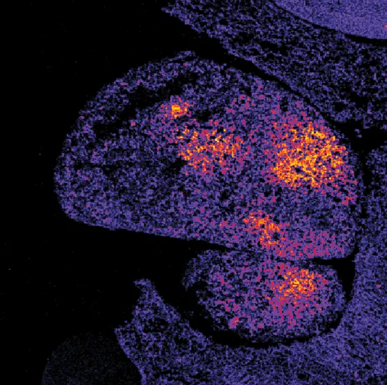
The DKFZ researchers led by Dick have now been able to solve this problem. Using a special combination of cold and chemical treatment, they were able to permanently lock the biosensor's state in the obtained tissue. Thus, they were able to obtain an image of the spatial distribution of oxidants in a fixed tissue section that corresponds to the one in the living organism.
In their publication, the investigators present the examples of oxidant distribution in a growing tumor, the response of the liver to inflammation, and the response of muscle fiber to hunger. The DKFZ researchers now plan to use the new method to study the effect of diseases and substances on the distribution of oxidants in the whole body.
In a second study, the scientists in Dick's group set out to further increase the sensitivity of the biosensors. This would enable them to observe subtle metabolism-related changes in the production of oxidants that occur, for example, as a result of dietary changes or physical activity.
Dick's team has now developed the first biosensor based on so-called peroxiredoxins, the proteins with the highest known sensitivity to hydrogen peroxide. The new probes respond with unprecedented sensitivity to the smallest increase or decrease in oxidant levels. When the investigators first tested the new probes in yeast cells, they were even able to monitor the movement of oxidants between individual structures within the cell.
Their next goal is to combine the two new developments. “We now also want to optimize the peroxiredoxin-based biosensors for mammalian cells and then place them into the genome of mice," explains Leticia Roma, who was a key researcher in the mouse study. “Combined with the possibility to analyze fixed tissue sections, we will then also be able to investigate whether minimal changes in oxidant production are linked to the development of metabolic diseases."
Fujikawa Y, Roma LP, Sobotta MC, Rose AJ, Diaz MB, Locatelli G, Breckwoldt MO, Misgeld T, Kerschensteiner M, Herzig S, Muller-Decker K and Dick TP (2016) Mouse redox histology using genetically encoded probes. Science Signaling 2016, DOI: 10.1126/scisignal.aad3895
Morgan B, Van Laer K, Owusu T, Ezerina D, Pastor-Flores D, Amphonsah P, Tursch A and Dick TP (2016) Real-time monitoring of basal H2O2 levels with peroxiredoxin-based probes. Nature Chemical Biology 2016, DOI: 10.1038/NCHEMBIO.2067


