Magnetic resonance imaging (MRI) is a method that is typically used to produce cross-section pictures of structures in the body’s soft tissues, such as organs. When exposed to a strong, artificially produced magnetic field, some of the hydrogen atoms in body tissues line up in a parallel state; when they are then exposed to radio frequency waves, they begin to oscillate. Depending on the structure and water content of the tissue under investigation, the hydrogen atoms that have thus been excited emit different signals; these are used to create a cross-section image through computational methods. The hydrogen atoms subsequently return to their original, unordered state. Due to the properties of tissues and atoms, the conventional technique can align and quantify only a very small portion of the hydrogen atoms – one in seven billion. The remaining atoms are invisible in MRI. Clinical MRI systems use very expensive special magnets to generate a magnetic field that is 100,000 times stronger and thus increases the alignment of hydrogen atoms, but this still produces an imagine of a few out of every million hydrogen atoms. More than 99,999 % remain invisible in MRI scans.
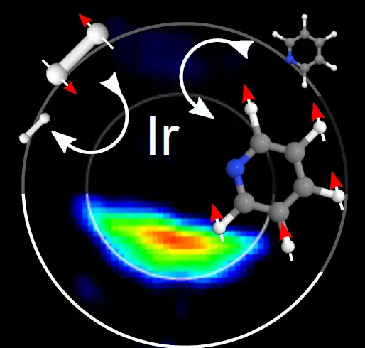
This prompted Hövener and his colleagues from the University Radiological Hospital in Freiburg, Germany, to take a different approach to enhancing the MRI signal. They used a method called hyperpolarization, which causes a magnetic alignment of a much greater proportion of the hydrogen atoms. The method also overcomes problems of previous attempts to employ hyperpolarization, namely that each atom could be polarized only once. The MR image itself destroys its alignment, making it impossible to obtain multiple images. To avoid this problem, the researchers from Freiburg and their colleagues from the Centre for Hyperpolarisation in York, U.K., used a form of hydrogen called parahydrogen, whose nuclei are in a specific quantum state. Parahydrogen can magnetically align other molecules by means of a chemical reaction; it can do so over and over in an appropriate magnetic field. This continuous polarization effect, based on previous work by researchers from York and Freiburg, is available for an unlimited time; it recovers after every measurement and thus facilitates repeated MR images. The signal thus emitted is 100 times stronger than in current MRI systems, even in a very low magnetic field, as can be produced, for example, using a simple battery.
“It is exciting to investigate this novel physical effect," says Hövener, who undertakes research at the Medical Physics Department of Diagnostic Radiology at the University Medical Center Freiburg and is a member of the German Consortium for Translational Cancer Research. He foresees numerous applications in chemistry and molecular biology. So far the scientists’ experiments have been restricted to the test tube, but tests in cell cultures and animal models will be the next step. As a long-term goal, Hövener hopes to exploit continuous hyperpolarization in biomedical research: “Hydrogen gas seems to be well tolerated by humans. Medical diagnosis may benefit tremendously from its use, but it is still a long way to get there." He thinks that possible applications in the future may include low-cost MRI systems to be used in medical screening as well as portable MRI scanners for on-site diagnoses.
Hövener J.-B., Schwaderlapp N., Lickert T., Duckett S.B., Mewis R.E., Highton L.A.R., Kenny S.M., Green G.G.R., Leibfritz D., Korvink J.G., Hennig J., von Elverfeldt D. A hyperpolarized equilibrium for magnetic resonance. Nature Communications, 2013.doi: 10.1038/ncomms3946


