Radiology
- Imaging and Radiooncology

Prof. Dr. Heinz-Peter Schlemmer
Leitung Abteilung Radiologie
The Division of Radiology at the German Cancer Research Center (DKFZ) is nationally and internationally recognized for its leading role in the development and application of advanced imaging technologies in oncology. Our work focuses on the early detection and assessment of tumor aggressiveness as well as the planning and evaluation of individualized therapies.

Welcome to the Division of Radiology

Our Commitment to Patients and Research
The well-being of our patients is at the heart of everything we do. In our research, we combine compassionate and professional care with the use of state-of-the-art imaging techniques to ensure precise, reliable, and patient-centered early detection and diagnosis at the highest level.
Innovative research and interdisciplinary collaboration
We combine medical and scientific expertise through close collaboration in the research focus area “Imaging and Radiation Oncology.” In this way, we continuously develop innovative imaging techniques and translate them into clinical practice.
Together with our partners, we are working to further develop key imaging technologies for cancer diagnostics:
- Sonography (ultrasound)
- Photon-counting computed tomography (CT)
- Magnetic resonance imaging (MRI) at various field strengths
- Positron emission tomography (PET) in combination with MRI (PET/MRI)
- Development and application of artificial intelligence
With the goal of further refining and personalizing early detection, diagnosis, and treatment planning, these methods are continuously refined in collaboration with our partners and evaluated and optimized within the framework of clinical trials.
Strong partnerships
Our research is conducted in close collaboration with leading national and international partners:
- Divisions and research groups at the DKFZ, particularly the Imaging and Radiation Oncology Research Topic
- Heidelberg and Mannheim University Hospitals
- National Center for Tumor Diseases (NCT) Heidelberg and the national NCT network with its other locations
- Research institutions of the Helmholtz Association and other renowned institutions
- Renowned national and international hospitals as well as international networks
- Medical technology and pharmaceutical industry partners
Our Mission
We develop innovative imaging methods for more precise, minimally invasive early detection, diagnosis, and personalized patient care.
Current Projects and Studies

EUCAIM (European Federation for Cancer Images) is establishing a European infrastructure for standardized cancer image data. The goal is to make radiological datasets available across Europe for research, AI development, and personalized cancer medicine, and to integrate them into the European Health Data Space (EHDS) in the long term.

4-IN-THE-LUNG-RUN (4ITLR) is a European multicenter study on the early detection of lung cancer using low-dose CT in high-risk groups. The goal is to develop personalized screening strategies with optimized screening intervals, reduced radiation exposure, and improved risk stratification in order to detect lung cancer earlier and make screening programs more efficient and safer in the long term.

The Li-Fraumeni syndrome whole-body MRI screening program investigates the use of radiation-free imaging for the early detection of cancer in individuals with a hereditary increased risk of cancer. The goal is to detect tumors as early as possible, improve preventive care, and establish low-burden screening strategies for affected patients.

HEROES-AYA investigates the heterogeneity, evolution, and treatment resistance of fusion-driven sarcomas in adolescents and young adults. Through multi-omics, digital pathology, and imaging, the project aims to better understand resistance mechanisms and develop new therapeutic approaches.
PROBASE is a prospective study on risk-adapted early detection of prostate cancer. The goal is to reduce overdiagnosis through PSA-based screening strategies and modern imaging, while simultaneously detecting clinically significant tumors at an early stage. The study aims to contribute to personalized and more efficient prostate cancer screening.

PUMA (Pulmonary MR-guided Online Adaptive Radiotherapy) is a multicenter clinical trial investigating MRI-guided adaptive radiotherapy for patients with locally advanced non-small cell lung cancer (NSCLC). The study aims to evaluate the feasibility and safety of daily anatomy-adapted radiation treatment in order to better spare healthy tissues while improving tumor control. The results will provide the foundation for future trials aimed at advancing personalized radiotherapy strategies.
Radiology Photo Gallery
-
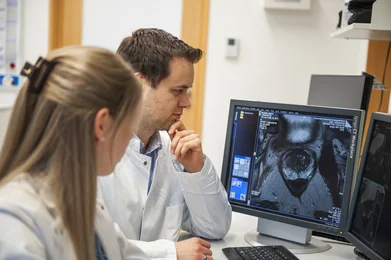
Diagnostik Radiologie -

Diagnostik Radiologie -

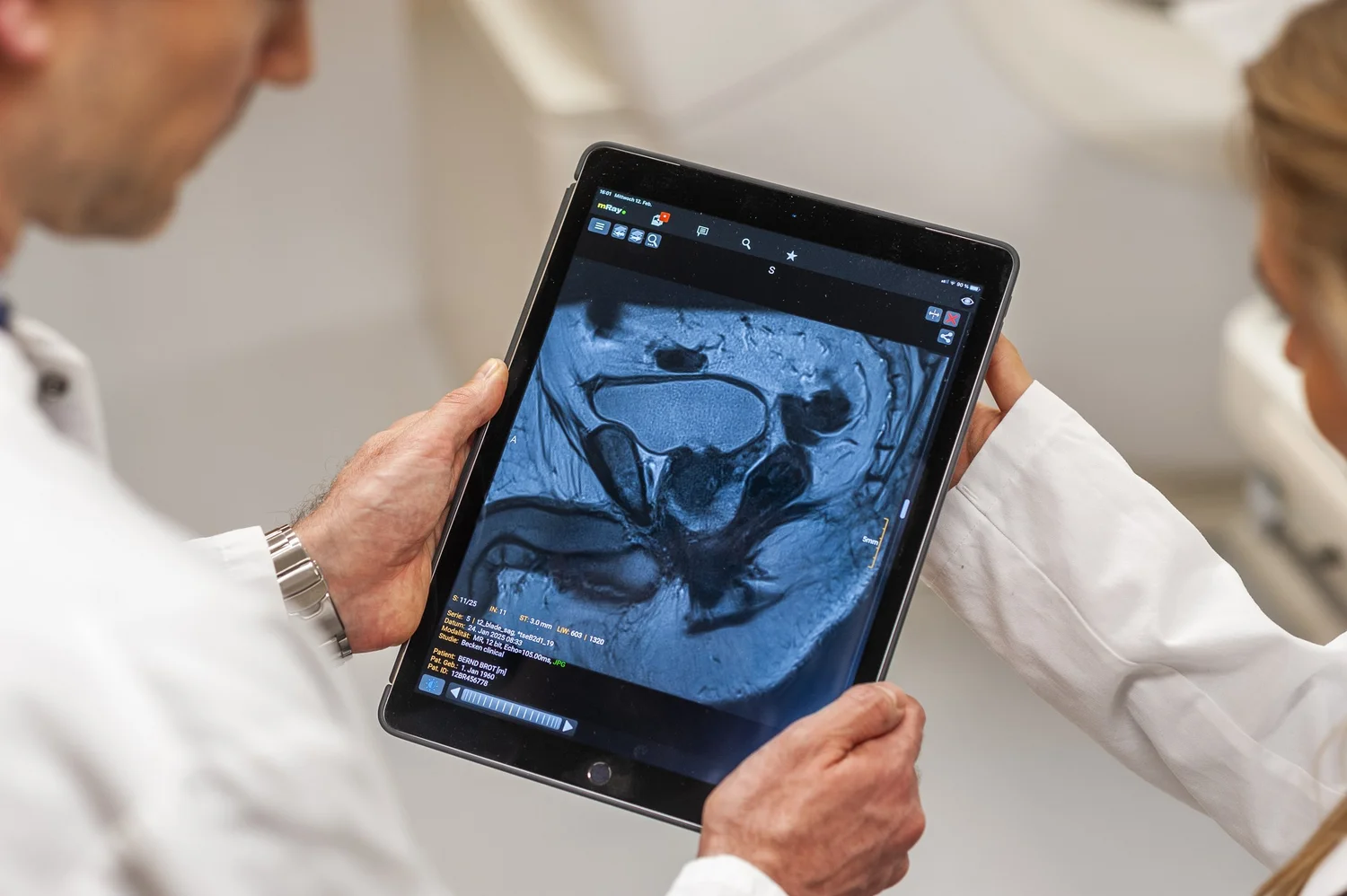
Prostata Diagnostik Radiologie -
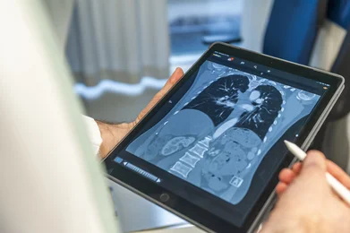
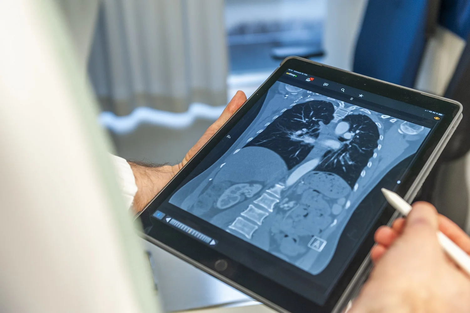
Thoraxdiagnostik Radiologie -
 im NCT")
Aera 1.5T (Siemens) im NCT -

Workplace for Radiology Medical Technologists at the NCT -

Registration for patients at the NCT -
 at the NCT")
Prisma 3T (Siemens) at the NCT -
 at the REZ")
NAEOMTOM Alpha. Peak (Siemens) at the REZ -
 at the REZ")
3T MR/PET hybrid system (mrBiograph, Siemens) at the REZ -

Registration for patients at the REZ -
")
Termin- und Informationsportal Radiologie (TIP) -

CT -
 at the REZ")
NAEOMTOM Alpha. Peak (Siemens) at the REZ


 im NCT")


 at the NCT")
 at the REZ")
 at the REZ")

")
 at the REZ")
Get in touch with us

