Cookie Settings

We use cookies to optimize our website. These include cookies that are necessary for the operation of the site, as well as those that are only used for anonymous statistic. You can decide for yourself which categories you want to allow. Further information can be found in our data privacy protection .

Essential

These cookies are necessary to run the core functionalities of this website and cannot be disabled.

Name Webedition CMS
Purpose This cookie is required by the CMS (Content Management System) Webedition for the system to function correctly. Typically, this cookie is deleted when the browser is closed.
Name econda
Purpose Session cookie emos_jcsid for the web analysis software econda. This runs in the “anonymized measurement” mode. There is no personal reference. As soon as the user leaves the site, tracking is ended and all data in the browser are automatically deleted.
Statistics

These cookies help us understand how visitors interact with our website by collecting and analyzing information anonymously. Depending on the tool, one or more cookies are set by the provider.

Name econda
Purpose Statistics
External media

Content from external media platforms is blocked by default. If cookies from external media are accepted, access to this content no longer requires manual consent.

Name YouTube
Purpose Show YouTube content
Name Twitter
Purpose activate Twitter Feeds

Researchers discover a possible reason for drug resistance in breast tumors

No. 33c3 | 20/07/2015 | by Koh

HER2 membrane proteins play a special role in certain types of breast cancer: amplified levels of HER2 drive unrestricted cell growth. HER2-tailored antibody-based therapeutics aim to prevent cancer cell growth. However, two-thirds of HER2 positive breast cancer patients develop resistance against HER2-targeting drugs. The reason for this is not yet understood. Researchers now found out, that HER2 dimers appeared to be absent from a small sub-population of resting SKBR3 breast cancer cells. This small subpopulation may have self-renewing properties that are resistant to HER2-antibody therapy and thus able to seed new tumor growth.

Single molecules and dimers of the HER2-receptor can be distinguished with Liquid STEM Microscopy
© Leibniz Institut für neue Materialien

For their studies researchers from the INM – Leibniz-Institute for New Materials, Saarbrücken and from the German Cancer Research Center (DKFZ) in Heidelberg used a new electron microscopy method called Liquid STEM. It allows nanoscale studies of intact cells in their native liquid environment.

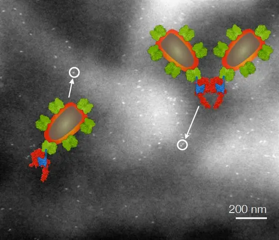
The scientists have studied the local variations of HER2 membrane protein and of its dimers. HER2 is a member of the human epidermal growth factor receptor (EGFR) family. These family members trigger cell growth signals, when two of the membrane proteins are bound into a protein complex (dimerization). This happens usually after the binding of a small protein, the epidermal growth factor, which circulates in the blood stream and serves as communicator to transmit signals that regulate cell growth. HER2 is special in the sense that it does not need the growth factor protein in order to form dimers. It is thus capable of triggering cell growth without external regulation. In certain types of breast cancer, amplified levels of HER2 and its dimerization are known to drive unrestricted cell growth. HER2-tailored antibody-based therapeutics entered clinical practice more than a decade ago. These drugs aim to prevent cell growth triggered by HER2 homo- and/or heterodimerization.

“We found out, that HER2 dimers appeared to be absent from a small sub-population of resting SKBR3 cells. Could such cells survive the therapy and then develop into a drug resistant cancer at a later stage? It thus seems to be of key significance to study this sub-population of cells with exceptional phenotype,” says Niels de Jonge, head of the Innovative Electron Microscopy group.

HER2 dimerization processes were thus far mostly studied on the basis of cell population averages, for example, with biochemical methods using pooled cell material, and information about the localization of HER2 dimerization was lacking. Therefore, the researchers around de Jonge pioneered the electron microscopy method Liquid STEM to imaging these receptors on cancer cells. The cells were examined on a microchip placed in the electron microscope, and remained intact and in liquid. “Specimens cannot be studied in liquid with traditional electron microscopy”, explains Professor de Jonge. “Cells are typically studied in dry state via thin sectioning of solid dried plastic embedded or frozen material. The role of HER proteins is a "hot" topic in cancer research but despite large research efforts using a wide range of techniques over the past decades this important information was not unveiled before. Our novel findings were obtained as a direct consequence of the high spatial resolution of Liquid STEM combined with its capability to study many intact cells in liquid,” says de Jonge.

D. B. Peckys, U. Korf, N. de Jonge: Local variations of HER2 dimerization in breast cancer cells discovered by correlative fluorescence and liquid electron microscopy. Science Advances, 2015, DOI: 10.1126/sciadv.1500165 

With more than 3,000 employees, the German Cancer Research Center (Deutsches Krebsforschungszentrum, DKFZ) is Germany’s largest biomedical research institute. DKFZ scientists identify cancer risk factors, investigate how cancer progresses and develop new cancer prevention strategies. They are also developing new methods to diagnose tumors more precisely and treat cancer patients more successfully. The DKFZ's Cancer Information Service (KID) provides patients, interested citizens and experts with individual answers to questions relating to cancer.

To transfer promising approaches from cancer research to the clinic and thus improve the prognosis of cancer patients, the DKFZ cooperates with excellent research institutions and university hospitals throughout Germany:

  • National Center for Tumor Diseases (NCT, 6 sites)
  • German Cancer Consortium (DKTK, 8 sites)
  • Hopp Children's Cancer Center (KiTZ) Heidelberg
  • Helmholtz Institute for Translational Oncology (HI-TRON Mainz) - A Helmholtz Institute of the DKFZ
  • DKFZ-Hector Cancer Institute at the University Medical Center Mannheim
  • National Cancer Prevention Center (jointly with German Cancer Aid)
The DKFZ is 90 percent financed by the Federal Ministry of Education and Research and 10 percent by the state of Baden-Württemberg. The DKFZ is a member of the Helmholtz Association of German Research Centers.

RSS-Feed

Subscribe to our RSS-Feed.

to top
powered by webEdition CMS