Magnetic resonance imaging, or MRI for short, provides detailed images of the inside of the body. It is an indispensable procedure for the diagnosis and treatment planning of many types of cancer, and it does not expose the body to radiation because it uses magnetic fields and low-energy radio waves. Pathological changes, such as tumors, can be clearly distinguished from healthy tissue in MRI images.
The element gadolinium provides the necessary contrast in MRI images; it is safely enclosed in the contrast agents within a so-called chelate molecule, much like a molecular cage. Through its magnetic properties, gadolinium interacts with water molecules in the body inside the MRI scanner. Certain tissues with a high water content thus appear brighter in the images, which creates contrast in the imaging.
Gadolinium-based contrast agents are used in just under half of all MRI scans. After the scan, these are excreted via the kidneys in the urine and end up in wastewater. There, gadolinium remains bound within its chelate cage in the long term. “Nevertheless, we see an urgent need to lay the foundations for new and sensitive analytical methods that will enable us to track gadolinium in the environment – not only from contrast agents but also from industrial processes,” explains Leif Schröder, Head of the Department of Translational Molecular Imaging at the DKFZ, who led the study.
Measuring Gadolinium Down to the Micromolar Range
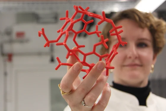
The scientists combined two effects. Firstly, they used hyperpolarized xenon, a strongly magnetized noble gas enclosed within a different type of cage, which produces signals in MRI. Here, the nuclear spin of the xenon acts as a quantum sensor and produces two distinct signals: one for the xenon bound within the cage molecules and one for the free xenon in the solution. Secondly, gadolinium displaces the xenon from its cage, causing the corresponding xenon signal to diminish and eventually disappear altogether.
“From the reduction in the xenon signal, we were able to determine the concentration of gadolinium outside the cage,” says Hannah Gerbeth, lead author of the study. The DKFZ research team found that gadolinium concentrations in the micromolar range can be measured. This makes the cage molecule a highly sensitive sensor for gadolinium under the conditions of the experiment. “This discovery shows that the unique properties of the interaction between gadolinium, xenon and the cage enable more sensitive detection than was possible in previous research using MRI,” says Leif Schröder.
The method for distinguishing free gadolinium from chelated forms could help improve its clinical application and the detection of gadolinium-containing substances in the environment. In future, the scientists plan to test the sensor’s suitability for measuring gadolinium in other concentration ranges as well. They also intend to investigate whether other ions are selectively bound by the cage and could therefore interfere with the gadolinium sensor.
Original Publication
Detection of Gd³⁺ via HyperCEST NMR in a Competition Assay Using Cryptophane-A Monoacid. Sens. Diagn. (2026) 5 (10): 1195–1203, doi.org/10.1039/d6sd00053c.


