Macromolecular organisation of striated muscle: Sarcomer from murine m. soleus. Insert: cross-section of acto-myosin setup (thick myosin- and thin actin-fibers).
Services
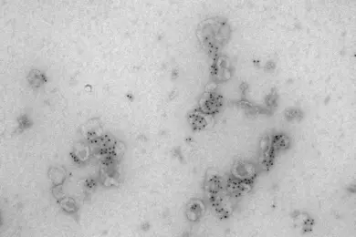
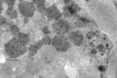
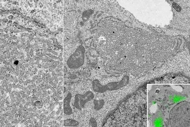
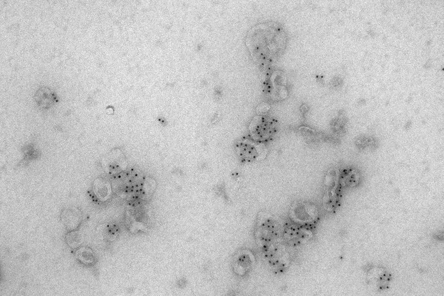
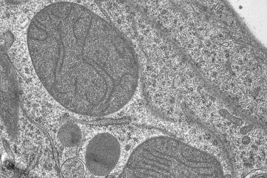
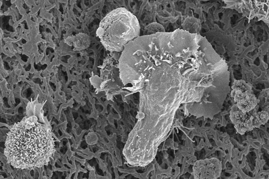
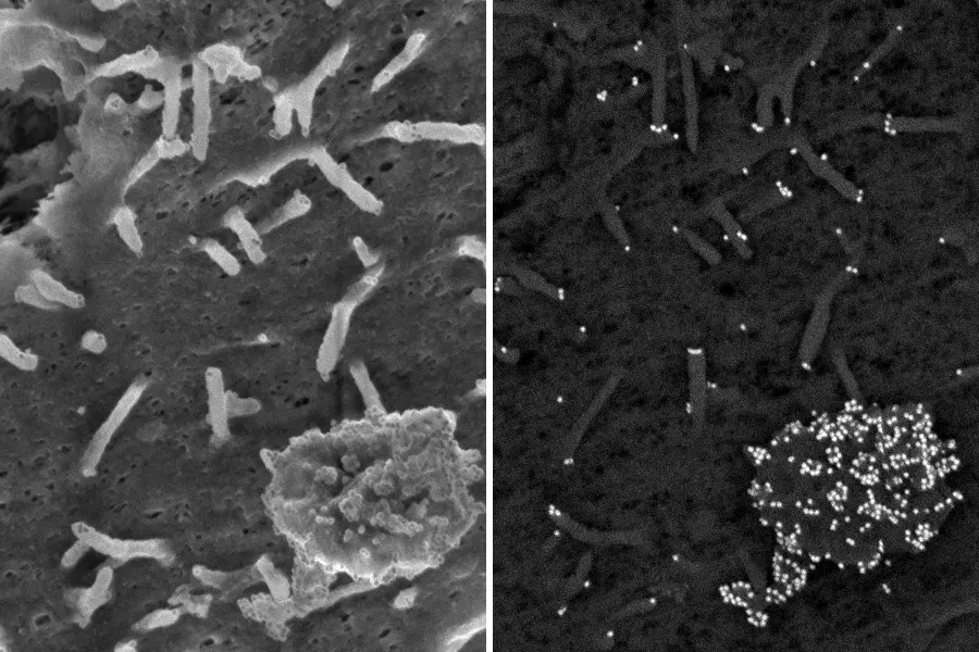
Since 2026 we jointly collaborate with the EM-Facility of Heidelberg University. Our service encompasses the design of experimental formats, sample preparation (fixation, embedding, sectioning, contrasting, labelling, carriers, etc.), microscopy, and pre-processing of images. Together with all image data, customers receive the detailed protocol and a report if appropriate. Our portfolio includes negative staining particle-TEM (quality-check of VLP- and EV-preparations), ultrathin sectioning resin-TEM (ultrastructural phenotyping of cells and tissues), as well as SEM (shapes and surface reliefes of cells, organoids and their substrates). Immuno-gold approaches furthermore allow allocation of molecular identity to structures of interest, and CLEM (Correlative Light and Electron Microscopy) to spot sites of interest by fluorescence microscopy. Our collaboration with the EMCF-Heidelberg adds capabilities in 3D approaches (TEM-tomography, 3D-SEM) and µCT. To start a project, contact us by mail or phone to arrange an appointment to clarify the scientific question and the experimental design.
As projects are unique, with respect to sample-formats and structural features of interest, we routinely invest into the development of protocols for sample preparation and imaging.