Neuroimmunologie und Hirntumorimmunologie
- Immunologie, Infektion und Krebs
- Klinische Kooperationseinheit

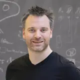
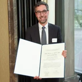
Prof. Dr. med. Michael Platten
Department Head
„Wir entwickeln innovative personalisierte Immuntherapien für Patient*innen mit Hirntumoren und überprüfen und verbessern diese in klinischen Studien."
Unsere Forschung

Das Zentralnervensystem (ZNS) wird als ein immunprivilegiertes Organ betrachtet, in dem Immunantworten trotz der Blut-Hirn-Schranke über einen intensiven Austausch mit dem peripheren Immunsystem streng kontrolliert werden. Unsere Gruppe entwickelt und verbessert immuntherapeutische Ansätze zur Bekämpfung von Hirntumoren mithilfe der Analyse molekularer Mechanismen der Immunsuppression und indem wir neuartige immuntherapeutische Behandlungsmodalitäten nutzen. Unsere Expertise liegt in umfassender zellulärer und auf Bildgebung basierender Analyse der Tumormikromilieus, Transcriptomics, sowie Profiling und Nutzung von Immunrezeptoren, sowohl in Mausmodellen und anhand von klinischen Proben. Unser zentraler Fokus ist die klinische Translation.
In den letzten Jahren haben wir immunsuppressive Funktionen und Mechanismen zweier von Hirntumoren produzierter Schlüssel-Metabolite identifiziert. Unsere Entdeckung, dass aus TDO entstandeneTryptophanmetabolite (Kynurenine) Tumorwachstum und Immunsuppression durch die Aktivierung des Arylhydrocarbonrezeptors (AhR) vermitteln, ermöglichte die Nutzung neuer therapeutischer Zielstrukturen und implizierte weitere Fragen, die wir aktuell in Tumormodellen adressieren. Ein zentrales Ziel ist die Identifizierung von Substanzen, die in den Tryptophankatabolismus eingreifen, um so potentielle Therapeutika für maligne Gliome zu entwickeln.
Weiterhin haben wir gezeigt, dass 2-Hydroxyglutarat, welches durch IDH-Mutationen von Gliomzellen produziert wird, direkt adaptive zelluläre Immunantworten, aber auch das inerte Immunsystem im Mikromilieu des Hirntumors inhibiert. Diese Erkenntnisse ebnen den Weg zu neuartigen Konzepten der immuntherapeutischen Kombinationstherapie, die wir momentan in präklinischen Gliommodellen untersuchen und in ihre klinische Translation begleiten.
Die Entwicklung neuartiger Zielantigene und T-Zellrezeptoren für die zielgerichtete Immuntherapie von Hirntumoren wurde in klinische Studien umgesetzt um die therapeutische Relevanz von mutationsspezifischen Vakzinen gegen klonale Hirntumor „Driver“ Mutationen zu demonstrieren. Laufende Projekte sind nun auf die Identifikation weiterer mutierter Antigene für die spezifische Immuntherapie und auf die Entdeckung spezifischer T-Zellrezeptoren (TZR) für die transgene T-Zelltherapie für Gliompatienten fokussiert. Unser Fokus liegt hierbei weiterhin bei CD4+ T-Helferzellen, die lange Zeit bezüglich ihrer Rolle bei der Immuntherapie vernachlässigt wurden. Hierfür haben wir einen Arbeitsablauf zur Entwicklung einer patientenspezifischen zielgerichteten Immuntherapie für Patienten mit Gliomen auf der Basis von Mutanomanalysen und der Identifikation von TZR, sowie ein MHC-humanisiertes murines Gliommodell zur präklinischen Evaluation entwickelt.
Bioinformatische Selektion reaktiver, in den Tumor infiltrierender T-Zellen zur TZR-Identifikation, basierend auf moderner Hochdurchsatz-Einzelzell-RNA-Sequenzierung, Einzelzell-VDJ-Sequenzierung sowie TZR-beta deep Sequenzierung, treiben diese Prozesse an und fördern ihre Entwicklung.
Mitarbeiter
-
Prof. Dr. med. Michael Platten
Department Head
-
Belize Acharya
MD-Student
-
Dr. Dennis Alexander Agardy
PostDoc
-
Kuralay Aman
MD Student
-
Dr. Gabor Bakos
PostDoc
-
Dr. Theresa Bunse
Teamleader
-
Dr. Lukas Bunse
Teamleader
-

Luis Diel
Intern
-
Amelie Christina Dietsch
MD Student
-
Andreas Dobbelstein
MD Student
-
Fabian Edmeier
MD Student
-
Alexander Ernst
Technical Assistance IMU
-
Henrike Feldmann
PhD Student
-
Hannah Gelhaus
PhD Student
-
Subhajit Ghosh
PostDoc
-

Mehsoon Gilani
PhD Student
-
Niklas Graßl
PostDoc
-
Dr. Edward Green
Teamleader
-
Sofia Heras Valdivielso
Intern
-
Julian Hlawatsch
MD Student
-
Zeren Hu
MD Student
-
Lena Hönig
Secretary
-
Melissa Höpfner
Technical Assistance
-
Ingrid Hülsmeyer
Technical Assistance IMU
-
Gyuyeon Jang
Intern
-
Kristine Jähne
Technical Assistance
-
Simone Jünger
Technical Assistance IMU
-
Sara Kangani
Asisstance
-
Vahid Khaki Bakhtiarvand
PhD Student
-

Philipp Koopmann
MD Student
-
Dr. Tom Niklas Kuhn
PostDoc
-
Ella Kuipers
Intern
-
Dr. Katharina Lindner
PostDoc
-
Claudia Maldonado Torres
Technical Assistance
-
Sarwar Mustafa
MD Student
-

Marie-Therese Neuhoff
PhD Student
-
David Palmero Canton
PhD Student
-
Alina Paul
PhD Student
-
Dr. Isabel Poschke
Teamleader
-
Fiona Regending
-
Fatemeh Rezazadeh
Intern
-
Marie-Yve Rudolph
-
Panthea Ruhdorfer
MD Student
-
Saskia Räuber
PostDoc
-
Dr. Katharina Sahm
Teamleader
-
Georgios Samaras
PhD Student
-
Khwab Sanghvi
PostDoc
-
Robin Seitz
Technical Assistance
-
Akanksha Shukla
Technical Assistance IMU
-

Aleksa Simic
Technical Assistance
-
Saskia Stange
PhD Student
-

Dr. Chin Leng Tan
PostDoc
-
Clara Tejido Dierssen
-
Ceyda Tozar
Student Assistant
-
Martin Uerlich
PhD Student
-

David Vonhören
PostDoc
-
Dr. Marie-Christine Wagner
Lab and project management
-
Franziska Widmayer
-
Julian Wiemer
-

Binghao Zhao
MD Student


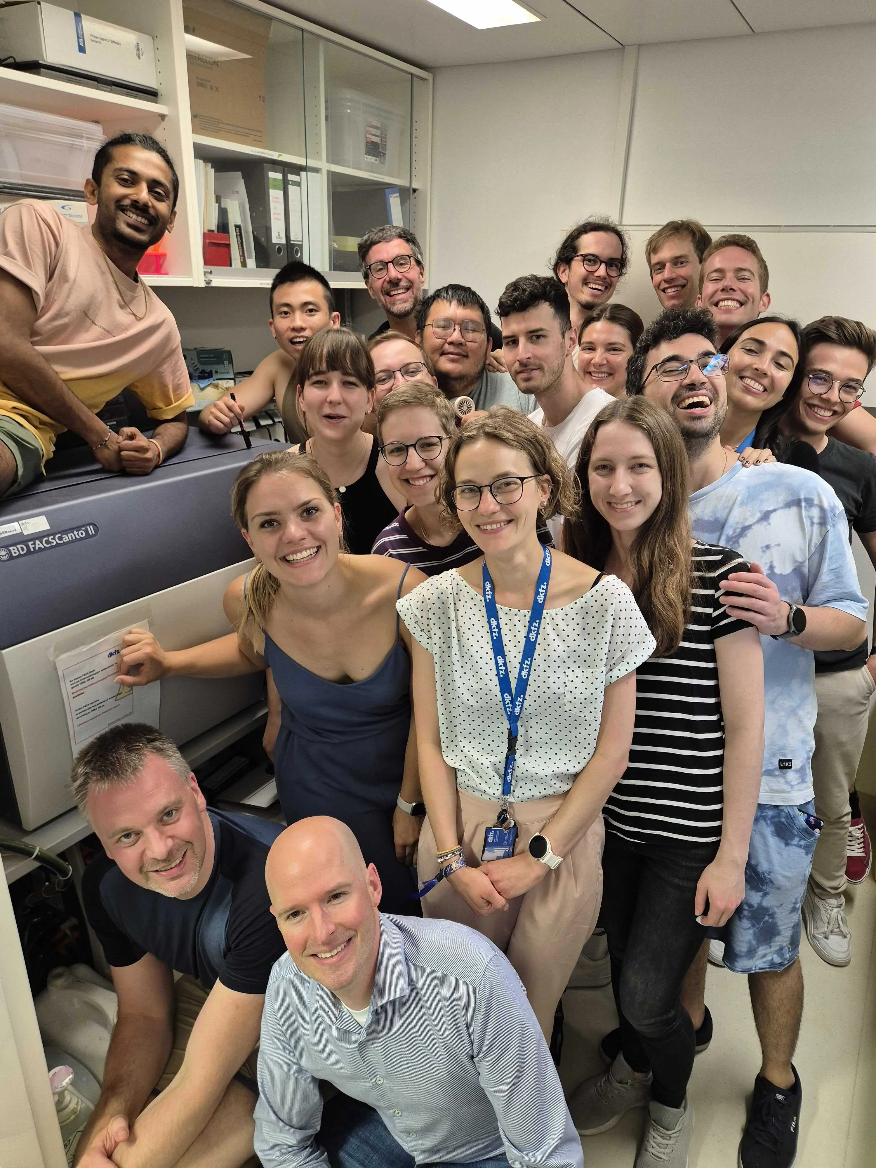
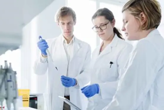
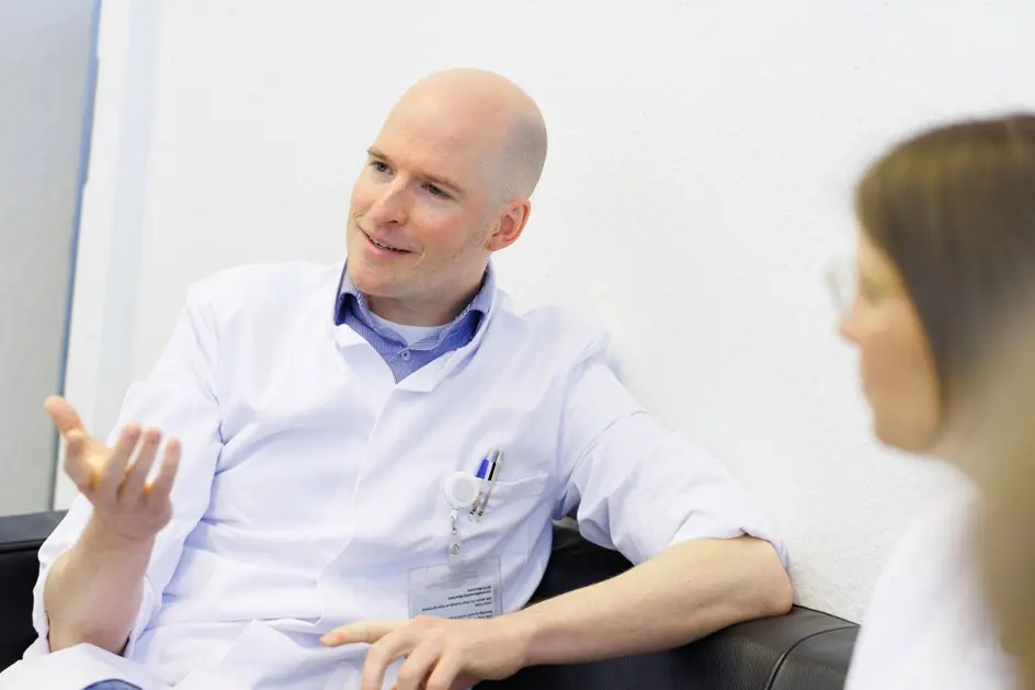
Bilder


Funding


Kontaktieren Sie uns
