Functional
Genome Analysis (B070)
Deutsches
Krebsforschungszentrum,
Im Neuenheimer Feld 580
D-69120
Heidelberg,
Germany. |
 |
.
.
..
FINISHED PROJECT:
Protein-based
microenvironmental communication in pancreatic ductal adenocarcinoma
We study the communication that is ongoing between the
various cell types in a tumour tissue, such as activated pancreatic
stellate cells and macrophages next to the tumour cells of pancreatic
ductal adenocarcinoma (PDAC). In particular, we analyse their
secretomes. Also, the effect on other cells is looked at. Cells
are grown in the different secretomes and are analysed with respect to
molecular and functional consequences. A graphical overview of the
process is shown to the right, presenting analyses done on pancreatic
stellate cells and the effect of their secretome on tumour cells.
Early, already published results are on the interaction of stellate and
tumour cells as well as the analysis of the tumour cell secretome.
Next to bilateral interactions, we have developed a system that allows
to study the interaction of several cell types simultaneously,
basically creating an environment under controlled conditions that
represents the cell composition of tumour tissue.
Marzoq et
al. (2019) Sci. Rep. 9,
5303. 
Mustafa
et
al.
(2017) Oncotarget
8,
11963-11976. 
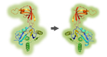
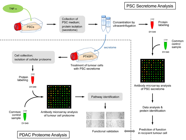
Figure legend: Scheme
of a typical overall experimental set-up of studying bilaterally the
interaction of two cell types: here, pancreatic stellate and tumour
cells. First,
the protein content of the secretome of activated pancreatic stellate cells (PSCs) was analysed and
predictions were made about the functional consequences, which the
secreted proteins would have in recipient tumour cells. Second, tumour
cells
were grown in media conditioned with secretome. The intracellular
proteome was
studied and used for functional predictions. The predictions from
secretome and intracellular proteome analyses were compared and
validated by
investigating the actual functional variations observed and by
identifying
relevant regulative factors.
|
|
|
.
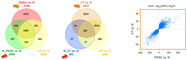
FINISHED PROJECT:
Transcript
variations in the wider peritumoral tissue environment of pancreatic
cancer
Transcriptional
profiling was performed on 452 RNA preparations isolated from various
types of
pancreatic tissue from tumour patients and healthy donors, with a
particular
focus on peritumoral samples. Pancreatic ductal adenocarcinomas (PDAC)
and
cystic tumours exhibited rather similar transcript patterns; in both
cases, about 5000 genes were differentially transcribed (see figure
below). In addition, all changes were identical in direction, up or
down (see figure, right panel). Not a single gene out of 5000 was found
to be up-regulated in one tumour but down-regulated in the
other.
..
PDAC and cystic tumours were most different in the non‐tumorous tissues surrounding them. As a matter of fact,
the
environment of
cystic tumours was transcriptionally nearly identical to normal
pancreas
tissue. In contrast, the tissue surrounding PDAC-tumours behaved a lot
like the
tumour itself -
indicating some kind of field defect - while showing far less molecular
resemblance to both chronic pancreatitis and healthy tissue. This
suggests that
major pathogenic differences between cystic and ductal tumours may
be due to
their cellular environment rather than the few variations within the
tumours.
..
Functionally,
a strikingly large number of autophagy‐related transcripts was changed in both PDAC and
its peritumoral tissue, but not in other pancreatic tumours. A
transcription
signature of 15 autophagy‐related genes was established that permits a
prognosis of survival with high accuracy and indicates the role of
autophagy in
tumour biology.
Bauer
et
al.
(2018) Int. J.
Cancer 142,
1010-1021.
|
|
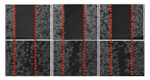
Figure legend: Tissue
specificity of mRNA level variations. For
each tissue type, the number of mRNAs is shown that were significantly
differentially expressed in comparison to normal pancreas tissue (N).
The
numbers in overlap regions stand for genes, regulated similarly in the
relevant
tissues. Left panel: Results are presented for PDAC, the related
peritumoral tissue
(N_PDAC) and chronic pancreatitis (CP), marked in red, green and
yellow,
respectively. Central panel: The panel presents the same
information for cystic tumours (TC;
brown),
the related peritumoral
tissue (N_CT; blue) and again chronic
pancreatitis (CP;
yellow). R ight panel: Correlation
in the direction of variation observed for CT versus N in comparison to
PDAC
versus N. Both axes represent the score shown above the panels, thus
focussing
on the most significant variations (shown in blue). Grey dots, mostly
close to
the centroid, represent insignificant changes.
|
.
|
..
FINISHED PROJECT:
Molecular signatures associated with tumor-specific
immune response in melanoma patients treated with dendritic cell-based
immunotherapy
We had previously shown that
autologous dendritic cells (DCs) loaded with an allogeneic heat shock
conditioned melanoma cell-derived lysate, called TRIMEL, induce
T-cell-mediated
immune responses in stage IV melanoma patients. Importantly, a positive
delayed-type
hypersensitivity (DTH) reaction against TRIMEL after vaccination
correlated
with patients prolonged survival. Furthermore, we observed that DTH
reaction
was associated with a differential response pattern reflected in the
presence
of distinct cell subpopulations in peripheral blood. Detected
variations in
patient responses encouraged molecular studies aimed to identify gene
expression profiles induced after vaccination in treated patients,
allowing the
identification of new molecular predictive markers. Gene expression
patterns
were analysed globally during vaccination, and some of them confirmed
in the
total leukocyte population of a representative group of responder and
non-responder patients. Seventeen genes overexpressed in responder
patients after
vaccination respect to non-responders were identified, of which ten
were linked
to immune responses and five related to cell cycle control and signal
transduction. In immunological responder patients, increased protein
levels of
the chemokine receptor CXCR4 and the Fc-receptor CD32 were observed on
cell
membranes of CD8+ T and B cells and the monocyte population,
respectively,
confirming gene expression results. Our study contributes to finding
molecular
markers associated with clinical outcome and better understanding of
clinically
relevant immunological responses induced by anti-tumour DC-vaccines.
rr
García et al. (2018) Oncotraget 9,
17014-17027. |
.
..
FINISHED PROJECT:
Melanoma
microRNA trafficking controls tumour primary niche formation


r
Melanoma
originates in the epidermis and becomes
metastatic after invasion into the dermis. Prior interactions between
melanoma
cells and dermis are poorly studied. Here, we show that melanoma cells
directly
affect the formation of the dermal tumour niche by microRNA trafficking
before
invasion.
..
Melanocytes, cells of melanoma origin, are specialized in
releasing
pigment vesicles, termed melanosomes. In melanoma in situ, we found melanosome
markers in distal fibroblasts before melanoma invasion. The melanosomes
carry
microRNAs into primary fibroblasts triggering changes, including
increased
proliferation, migration and pro-inflammatory gene expression, all
known
features of cancer-associated fibroblasts (CAFs). Specifically,
melanosomal
microRNA-211 directly targets IGF2R and leads to MAPK signalling
activation,
which reciprocally encourages melanoma growth. Melanosome release
inhibitor
prevented CAF formation. Since the first interaction of melanoma cells
with
blood vessels occurs in the dermis, our data suggest an opportunity to
block
melanoma invasion by preventing the formation of the dermal tumour
niche.
rr
Dror et al. (2016) Nature Cell Biol. 18,
1006-1017.
|
|
 |
..
..
FINISHED PROJECT:
Expansion of a BDCA1+CD14+
myeloid cell population in melanoma patients may attenuate the efficacy
of
dendritic cell vaccines.
The tumour microenvironment
is characterized by regulatory T cells, type II macrophages,
myeloid-derived
suppressor cells, and other immunosuppressive cells that promote
malignant
progression. Here we report the identification of a novel
BDCA1(+)CD14(+)
population of immunosuppressive myeloid cells that are expanded in
melanoma
patients and are present in dendritic cell-based vaccines, where they
suppress
CD4(+) T cells in an antigen-specific manner. Mechanistic
investigations showed
that BDCA1(+)CD14(+) cells expressed high levels of the immune
checkpoint
molecule PD-L1 to hinder T-cell proliferation. While this
BDCA1(+)CD14(+) cell
population expressed markers of both BDCA1(+) dendritic cells and
monocytes,
analyses of function, transcriptome, and proteome established their
unique
nature as exploited by tumours for immune escape. We
propose that targeting these cells may improve the efficacy of cancer
immunotherapy.
rr
Bakdash et al. (2016) Cancer Res. 76,
4332-4346. |
.
 |
|
FINISHED PROJECT:
Early
epigenetic down-regulation of microRNA-192 expression promotes
pancreatic cancer progression |
 |
r
Pancreatic
ductal adenocarcinoma (PDAC) is
characterized by very early metastasis, suggesting the hypothesis that
metastasis-associated changes may occur prior to actual tumor
formation. We
identified miR-192 as an epigenetically regulated suppressor gene with
predictive value in this disease. miR-192 was downregulated by promoter
methylation in both PDAC and chronic pancreatitis (CP), the latter of
which is
a major risk factor for development of PDAC. Functional studies in vitro and in vivo in mouse models of
PDAC showed that overexpression of
miR-192 was sufficient to reduce cell proliferation and invasion.
Mechanistic
analyses correlated changes in miR-192 promoter methylation and
expression with
epithelial-mesenchymal transition (EMT). Cell proliferation and
invasion were
linked to altered expression of the miR-192 target gene SERPINE1
that is encoding the protein plasminogen activator
inhibitor-1 (PAI-1), an established regulator
of these properties in PDAC cells. Notably, our data suggested that
invasive
capacity was altered even before neoplastic transformation occurred, as
triggered by miR-192 downregulation. Overall, our results highlighted a
role for
miR-192 in explaining the early metastatic behavior of PDAC and
suggested its
relevance as a target to develop for early diagnostics and therapy.
.
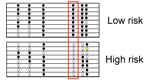
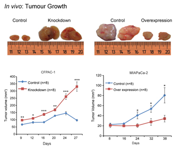
Figure legend.
Two cell lines, which express miR-192 at high level (CFPAC-1) or low
level (MIAPaCa-2) were transfected with constructs that suppressed or
increased miR-192 expression, respectively. Xenografted into mice, strong effects on
tumour growth were observed.
rr
Botla et al. (2016) Cancer Res. 76, 4149-4159. |
FINISHED PROJECT:
COST Action
Pancreatic Cancer
|
|
|
Large-scale international
collaboration is essential to decipher relevant information in the
context of
omics-scale interrogations in cancer research. This is even more
important for relatively
rare but very fatal diseases like pancreas cancer. The COST Action
Pancreatic
Cancer facilitated the collaboration of a broad range of
European
and
international research groups on pancreatic cancer in order to: (1)
integrate
knowledge and experience in a multidisciplinary way ‘from molecule via
cell to
society’, (2)
promote the application of uniform study tools and protocols, (3)
foster their
optimal use by early-stage researchers, (4) enhance the mobility and
training
of researchers, and (5) disseminate the results produced to society.
..
The Action developed novel
tools that improved our understanding of pancreatic cancer and its
prevention,
diagnosis and treatment. It also aimed at answering questions related
to
the tumours’
etiology, early detection, evidence-based and personalised treatment,
as well as to
aspects of health management. We aimed at attracting young scholars
across a
range of disciplines, who worked in collaboration with more
experienced
researchers. This enhanced active European participation in the
international research efforts on pancreatic cancer, with the objective
of
reducing disease mortality.
|
|


… |
Milne et al. (2014) Public Health Genomics
16, 305-312.
|
|
|
|
..
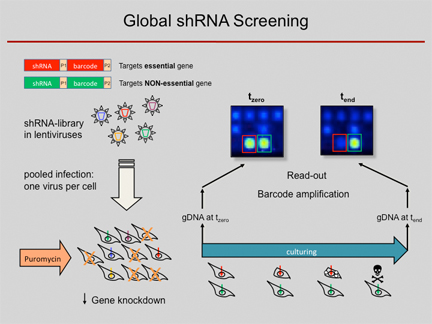
Scheme of genome-wide
shRNA knockdown experiments. Meanwhile, the read-out is done by
next-generation sequencing instead of microarray analysis as depicted.
|
|
FINISHED PROJECT:
Functional
screens by means of lentiviral shRNA libraries
RNA
interference (RNAi) has become a popular and important tool for the
analysis of gene
function.
Loss-of-function studies, commonly performed by transfection of short
interfering RNAs (siRNAs), have greatly facilitated functional analyses
of the
human transcriptome. However, there are major downsides to siRNA
experiments,
most importantly the transient inhibition of gene expression as well
as their
inefficient transfection into non-dividing cells. Overcoming
those
limitations, short hairpin RNA (shRNA)
expression
vectors are available, which
stably integrate into a target cell's genome via
retro-
or lentiviral gene transfer. Intracellular processing of shRNAs results
in
short duplex RNAs with siRNA-like properties. Viral integration ensures
a broad range of infectable target cell types and a stable
expression of specific shRNAs, resulting in the permanent reduction of
the
targeted gene product. Complex shRNA expression libraries
allow the targeted knockdown of
thousands
of different genes in a single experiment.
...
Using
such lentiviral vector
shRNA libraries and
initially barcode arrays and meanwhile next-generation sequencing analysis for decoding of the pooled RNAi screens, we are able to quantify the
abundance of
individual shRNAs and thus determine in a complex pool the number of
cells infected with an individual shRNA construct. We used the
technique to
predict anti-proliferative effects of individual shRNAs from pooled
negative
selection screens, for example, identified synthetic-lethal
activities toward combination
therapies, defined genes which
are required
for a stem-cell
like phenotype and found tumour suppressor
genes by in vivo studies.
...
Further
studies are under way, both for the elucidation of basic regulative
processes associated to cancer and for the identification of pathways
that are affected by particular drugs or compounds. In particular, we
use the technique for obtaining more detailed information
on the functional effects of particularly potentially druggable gene
products.
..
More recently, 259,900 CRISPR-Cas9 constructs have been
used for whole-genome analyses.
|
|
Wolf et al. (2014) Oncogene 33, 4273-4278. |
|
|
Fredebohm et al. (2013) J. Cell Sci. 126, 3380-3389. |
|
|
|
Böttcher et al. (2014) BMC Genomics 15,
158. |
|
|
Böttcher et al. (2010) BMC Genomics 11, 7. |
|
|
|
Wolf et al. (2013) Breast Cancer Res. 15, R109. |
|
|
Böttcher & Hoheisel (2010) Curr. Genom. 11, 162-167. |
|
|
|
|
a
FINISHED PROJECT:
Breast cancer
An
in vivo RNAi
screen
identifies SALL1 as a tumour
suppressor in human breast cancer with a role in CDH1 regulation
A
The gold standard for
determining the tumorigenic potential of human cancer cells is a
xenotransplantation into immunodeficient mice. Higher tumorigenicity of
cells
is associated with earlier tumour onset.
A
We employed
xenotransplantation to assess the tumorigenic potential of human breast
cancer
cells following RNAi-mediated inhibition of over 5,000 genes. We
identified sixteen
candidate tumor suppressors, one of which is the zinc finger
transcription
factor SALL1. Analysing this
particular molecule in more detail, we showed that inhibition of SALL1 correlated with reduced levels of CDH1,
an important contributor to epithelial-to-mesenchymal
(EMT) transition. Furthermore, SALL1
expression led to increased migration and more than twice as many cells
expressing a cancer stem cell signature. Also, SALL1
expression correlated with the survival of breast cancer
patients. These findings cast new light on a gene, which has previously
been
described to be relevant during embryogenesis, but not carcinogenesis.
A
Wolf et al. (2014) Oncogene 33, 4273-4278.
FINISHED PROJECT:
Breast cancer
A
mammosphere formation RNAi screen reveals genes that promote a breast
cancer stem-like phenotype
A
Breast cancer stem cells
are suspected to be responsible for tumour recurrence, metastasis
formation as
well as chemoresistance. Consequently, great efforts are made to
understand the
molecular mechanisms underlying cancer stem cell maintenance. In order
to study
these rare cells in vitro, they are
typically enriched via mammosphere culture. We developed a
mammosphere-based
negative selection shRNAi screening system suitable to analyse the
involvement
of thousands of genes in the survival of cells with cancer stem cell
properties. We used a sub-population with cancer stem cell properties
of cell
line SUM149 that were enriched in mammospheres. Identified
candidates were validated in mammosphere and soft agar colony formation
assays.
Further, we evaluated the influence of their expression on the stem
cell
sub-population. Also, the tumorigenic potential of SUM149 after up- or
down-regulation of candidates was examined by xenograft experiments.
A
Using this approach, Jak-STAT as well as cytokine
signalling were identified to be involved
in mammosphere formation. Furthermore, the autophagy regulator ATG4A
was found
to be essential for maintenance of the cancer stem cell sub-population and regulation of breast cancer cell
tumourigenicity in vivo.
A
Wolf et al. (2013) Breast Cancer Res. 15, R109.
|
FINISHED PROJECT:
Synthetic-lethal screens:
Depletion
of
Rad17 sensitises pancreatic cancer cells to gemcitabine
A
Chemotherapy
of advanced pancreatic cancer has mainly been gemcitabine-based, but
with only
limited effect. Recently, combination therapy that also targets
checkpoint
kinase 1 (CHK1) has become an attractive option. The central role of
CHK1 in
many DNA damage response pathways, however, may result in undesired
cytotoxicity in normal cells. We were searching for other target
molecules that
may be more specific and thus better suited for combination therapy.
A
A
negative
selection RNAi screen was performed, targeting over 10,000 genes. Genes
that
were found to be synthetically lethal with gemcitabine and whose
proteins are
acting upstream of CHK1 were characterised in more detail. The
inhibition of
RAD17 potentiated gemcitabine cytotoxicity particularly, leading to
forced
mitotic entry of cells arrested in S-phase by gemcitabine treatment,
resulting
in asymmetric DNA distribution during anaphase followed by DNA
fragmentation
and finally cell death by mitotic catastrophe.
A
Our
data suggest RAD17 for
gemcitabine combination therapy supplementing or complementing
inhibition of
checkpoint kinase 1. As opposed to CHK1, RAD17 knockdown by itself does
not
inhibit cell proliferation and does not lead to abnormal DNA
segregation,
suggesting a more specific action.
A
Fredebohm et al. (2013) J. Cell Sci. 126, 3380-3389.
|
....
FINISHED PROJECT:
NGFNplus
Translational Genome Research Network Pancreatic
Cancer |
|
...
 Genome
projects have
generated knowledge and technology with great potential to contribute
to the
understanding of the molecular pathogenesis in the pancreas and to
provide
molecular targets. However, even though multiple genome scale screening
approaches of pancreatic tumours and their preneoplastic lesions have
been
conducted, only few targets have reached the level of preclinical or
clinical
applications.
The PaCaNet
consortium was an
Integrated Genome Research Network comprising groups who (i) set the
standards
of clinical care and histopathology of pancreatic cancer and its
precursor
lesions, (ii) have pioneered the use of high-throughput genome
technology in
pancreatic research, (iii) have generated in-vitro and in-vivo models
of
the
disease and (iv) were among the first to transfer individual target
genes or
groups of target genes into preclinical and clinical applications.
These German
centers of excellence in pancreatic cancer research were joined by
genome
research groups and partners from the pharmaceutical industry in an
integrated
approach for an efficient characterisation and exploitation of genome
project
candidate genes for pancreatic cancer. The prime objective was to
foster
the
rapid development and transfer of novel genome-based, molecular
targeted
therapeutic and diagnostic approaches from basic research, over
preclinical
testing into clinical applications. The workplan
was conducted
at several levels of research: patforms:
data,
patients, resources, models; functional
characterisation in human in vitro and mouse in
vitro and in vivo models; preclinical
testing in mouse models of pancreatic cancer; and clinical
testing. Genome
projects have
generated knowledge and technology with great potential to contribute
to the
understanding of the molecular pathogenesis in the pancreas and to
provide
molecular targets. However, even though multiple genome scale screening
approaches of pancreatic tumours and their preneoplastic lesions have
been
conducted, only few targets have reached the level of preclinical or
clinical
applications.
The PaCaNet
consortium was an
Integrated Genome Research Network comprising groups who (i) set the
standards
of clinical care and histopathology of pancreatic cancer and its
precursor
lesions, (ii) have pioneered the use of high-throughput genome
technology in
pancreatic research, (iii) have generated in-vitro and in-vivo models
of
the
disease and (iv) were among the first to transfer individual target
genes or
groups of target genes into preclinical and clinical applications.
These German
centers of excellence in pancreatic cancer research were joined by
genome
research groups and partners from the pharmaceutical industry in an
integrated
approach for an efficient characterisation and exploitation of genome
project
candidate genes for pancreatic cancer. The prime objective was to
foster
the
rapid development and transfer of novel genome-based, molecular
targeted
therapeutic and diagnostic approaches from basic research, over
preclinical
testing into clinical applications. The workplan
was conducted
at several levels of research: patforms:
data,
patients, resources, models; functional
characterisation in human in vitro and mouse in
vitro and in vivo models; preclinical
testing in mouse models of pancreatic cancer; and clinical
testing.
|
|
.
|
FINISHED PROJECT:
Pancreatic cancer
susceptibility loci and their role in survival...
There are strong epidemiologic evidences indicating
that genetic common variability could be implicated in the risk
developing of
pancreatic cancer and various risk loci have been proposed. Genome-wide
association studies (GWAS) have been performed worldwide, and resulted
in
several loci associated with risk of pancreatic cancer.
In the context of the
PANcreatic Disease ReseArch (PANDoRA) consortium, coordinated by
Federico
Canzian (DKFZ), we replicated the associations found by others in two additional, independent populations (from Germany and the UK) and also evaluated the possible impact of
these SNPs on patient survival. For
the latter, we focused particularly on the ABO
locus. Moreover, we performed stratified analyses considering the tumor
stage
in order to verify whether genetic variability could be involved in the
disease
prognosis. Also, we attempted to replicate novel risk loci
identified in studies conducted in Japan and China in individuals of European descent.
|
|
|
|
|
Campa et al. (2013) Can.
Epidem. Biomarkers Prev. 22, 320-322. |
|
|
|
|
|
Rizzato et al.
(2013) Oncol. Report 29, 1637-1644. |
|
|
|
|
|
Rizzato et al. (2011) PLoS ONE
6,
e27921. |
|
|
|
|
|
|
.
.
FINISHED PROJECT:
Mutation spectrum
and prognostic significance of K-RAS
and CDKN2A in exocrine
pancreatic tumours ...
K-RAS mutations are major
factors involved in initiation and maintenance of pancreatic tumors.
The impact
of different mutations on patient survival has not been clearly
defined.
Therefore, we screened tumors from 171 pancreatic cancer patients for
mutations
in K-RAS, CDKN2A, BRAF
and GNAS genes. Mutations in K-RAS
were detected in 134 tumors, with
131 in codon 13 and 3 in codon 61. The GGT>GAT (G12D) was the most
frequent
mutation and present in 60% (80/134). Deletions and mutations in CDKN2A were detected in 43 tumors; GNAS
mutations were present in two
tumors.
Analysis showed that K-RAS mutations
were associated with
reduced patient survival in all sub-categories. Patients with malignant
exocrine tumors that had K-RAS
mutations showed a median survival of 17 months compared 30 months for
those
without mutations. Patients with a G12D mutation showed a median
survival of 16
months. Although the association of survival in pancreatic cancer
patients with CDKN2A aberrations in tumors was not
statistically significant, the sub-group of patients with concomitant K-RAS mutations and CDKN2A alterations
in tumors were associated with a median survival
of 13 months compared 30 months without mutation. Our results clearly
show an
association between mutational status and survival in pancreatic cancer
patients.
Rachakonda et al. (2013) PLoS ONE 8,
e60870. |
|
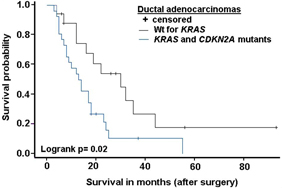
Kaplan-Meier
survival curves
showing difference in overall survival in PDAC patients with and
without
mutations.
|
|
FINISHED PROJECT:
Breast
Cancer
Epigenetically
de-regulated miR-375 is involved in a positive feedback loop with
Estrogen Receptor alpha in breast cancer cells
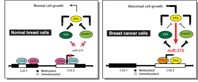

Breast cancer is the
leading cause of cancer death in women worldwide. Although it is a
heterogeneous disease, two-thirds of breast cancers share the common
feature of
being dependent on the presence and interaction of estrogen with the
nuclear
Estrogen Receptor α (ERα) protein. Approximately 70% of
invasive breast cancers express ERα in actively proliferating cells. It
has
become evident that ERα is up-regulated in luminal mammary epithelial
cells
during early stages of tumorigenesis and its overexpression is an
important
stimulatory factor for proliferation
of mammary cells, leading to cell division and eventually to tumor
development. The obvious role of ERα
signalling in orchestrating the expression of genes involved in
growth-related
pathways, has established ERα as an important therapeutic target in
breast
cancer treatment. However, our understanding of the molecular
mechanisms
underlying deregulation of this signaling pathway is scarce.
...
We identified a high
expression of microRNA 375 (miR-375) in ERα-positive cell lines.
miR-375
overexpression is mainly caused by the loss of epigenetic marks, such
as
H3K9me2 and local DNA hypomethylation, which, in turn, triggers the
dissociation of the transcriptional repressor CTCF from the promoter
and
enables interactions of ERα with regulatory regions of miR-375.
Inhibition of
miR-375 in ERα positive MCF-7 cells results in reduced ERα activation
and cell
proliferation. A combination of expression profiling from tumour
samples
and
miRNA target prediction identified RASD1 as a potential miR-375 target.
Our
findings show that miR-375 regulates RASD1 through targeting its 3'
UTR. In
addition, we demonstrated that RASD1 negatively regulates ERα
expression. Our
data indicate the existence of a positive regulation between ERα and
miR-375
and suggest new strategies for the treatment of ER-positive invasive
breast
tumours.
de Soza Rocha Simonini et
al. (2010) Cancer Res. 70, 9175-9184.
|
.
FINISHED PROJECT:
Pancreatic Cancer
Developing
novel molecular tools for the prevention
and diagnosis of pancreatic cancer
...

  |
While
project funding has ceased, the collaboration between many partners
involved in this consortium continues nevertheless.
The overall
aim of this EU
Framework Programme 6 Integrated Project was to make use of genetic
profiles of
pancreatic cancer and precursor lesions to improve the outcome of
pancreatic
cancer patients by providing novel and highly efficient molecular
diagnostic
tools. One of the major prerequisites in order to achieve this
ambitious aim was
an integrated multidisciplinary research approach, which enabled a
strong
interaction between technology, biology and medicine to translate
genome data
into practical, clinical applications.
The
consortium included molecular
biologists, bioinformaticians, pathologists, epidemiologists, molecular
oncologists, surgical and medical oncologists, radiologists and nuclear
medicine physicians. Since we expected to generate molecular diagnostic
tools
that will be ready for clinical applications, a
number of companies was involved that have a particular interest in
developing
molecular diagnostic tools, and one partner from pharmaceutical
industry.
|
The
scientific objectives of the project were defined in seven workpackages:
..
Level 1: Data, patients, resources:
WP1:
Epidemiology,
patients at risk, patient resources, coordinated
by J.P. Neoptolemos, W.
Greenhalf and N. Malats
WP2:
Molecular
alterations of preneoplastic lesions, early and advanced tumours:
genomic and
proteomic profiles, coordinated by
N. Lemoine & T. Jurcevic
...
Level
2: Development of novel molecular diagnostic tools
WP3:
RNA-based
diagnostics, coordinated by T.M.
Gress
WP4:
Proteome based diagnostics, coordinated
by E. Costello
and S. Hahn
WP5:
Epigenetics, coordinated by J.D.
Hoheisel
WP6:
Novel Molecular Imaging tools based on single proteins
identified in high-throughput approaches, coordinated by S. Hahn
...
Level 3: Clinical trials of
novel molecular diagnostic tools:
WP7:
Prospective clinical trials of novel molecular
diagnostic tools, coordinated by
J.P.
Neoptolemos
The
project used clinical samples such as serum, urine, fine needle
aspirates
and surgically resected materials of pancreatic cancer patients
collected in
large multinational European trials such as ESPAC or EUROPAC. During
the last
phase prospective clinical trials of novel diagnostic tools developed
in the
integrated project were designed and started.
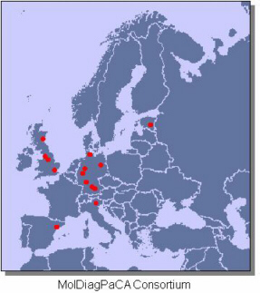
For
more information see MolDiagPaCa
webpage. |
|
 |
|
.
.
FINISHED
PROJECTS:
Identification
and characterisation of disease genes
by Representational Difference Analysis
|
 .
. .
. |
| Complementary
and parallel to
differential hybridisation techniques, Representational Difference
Analysis
(RDA) was employed as an alternative method for the detection of
differentially
expressed genes. The technique was adapted from the original protocol
of Hubank
and Schatz (1994) so that analyses are possible even with small amounts
of
starting material. In various projects, this technique was applied for
the
isolation of genes related with and potentially causative to diseases.
In
collaborations with the companies Merck, Hoffman la Roche and Knoll
(now
Abbott), analyses were carried out for the identification of
disease-related
genes on a wide variety of tissues. Large number of cancer-related
transcriptional differences were identified and some of
the relevant genes and gene products analysed in more detail. Also,
antibodies were generated on the basis of such studies and are being
used in
actual protein expression analyses. |
.
 . .
|
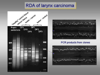
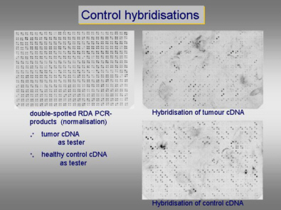
RDA was
performed on larynx
carcinoma tissue versus normal larynx tissue. Total difference products
were
cloned and and individula clones were picked. Redundancy of the library
was
minimised by iterative hybridisations of clones back to the library.
PCR-products were spotted on filters, with the clones representing
overexpression in normal and cancer tissue, respectively, being spotted
in
different orientation. Upon hybridisation of the respective starting
material,
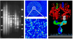
all but one signal were of the expected orientation. |
|
|
|
|
|
|
|
|
Schütz et al. (2006).J. Mol. Biol. 358, 997-1009. |
|
|
Frohme et
al..(2002) Biochem. Biophys. Res.
Commun. 293, 1377-1382. |
|
|
|
|
|
|
Steinberg et al. (2006).J. Periodontal. Res. 41, 426-446. |
|
|
Frohme et
al..(2000) Ann. N.Y. Acad. Sci. 910, 85-104. |
|
|
|
|
|
|
Frohme &
Hoheisel (2006).Cell Biology
3rd ed., Elsevier, 113-120. |
|
|
Geng et al..(1998) Biotechniques 25,
434-438. |
|
|
|
|
|
|
Malanchi et al..(2004).J. Virol. 78, 13769-13778. |
|
|
Gress et
al.
(1997) Genes Chrom.
Cancer 19, 97-103. |
|
|
|
|
|
|
Zubakov et al..(2003).FEBS Lett. 547, 51-57. |
|
|
-------------------------------------------------------------------------------------------------
other
publications and patents
|
|
|
|
|
|
|
Scheideler
& Hoheisel (2002) Screening 5/02,
22-25. |
|
|
|
|
|
|
|
|
|
|