Mammalian Cell Cycle Control Mechanisms Prof. Ingrid Hoffmann
Scientific Projects:
Mammalian Cell Cycle Control Mechanisms
Ongoing projects in the lab
1. Centrosome duplication, chromosomal instability and cancer
Failure to properly control centrosome number results in supernumerary centrosomes, which are frequently found in cancer cells. Centrosomes are small organelles that contain a pair of barrel-shaped centrioles surrounded by pericentriolar material, PCM. Centrioles duplicate once during the cell cycle to give rise to two mitotic spindle poles, each containing one old and one new centriole. Centrosome duplication must occur in coordination with other cell cycle events, including DNA synthesis.
Most human cancers exhibit centrosome duplication errors which might lead to aneuploidy and cancer formation. How centrioles are assembled and how their numbers are controlled within cells constitute long-standing unresolved questions.
Plk4, a polo-like kinase family member, is the key regulator of centriole duplication. We are studying the pathways underlying Plk4-induced centriole duplication in normal and malignant cells by identifying and characterizing substrates and regulators of Plk4.
 more
more
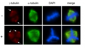
Figure 1: Overexpression of Plk4 leads to the formation of multipolar mitotic spindles. A tetracycline-inducible Plk4 U2OS cell line was generated. Plk4 expression was induced through removal of tetracycline from the medium. Red: centrosomal staining (marked with arrows) (γ-tubulin), green: microtubule staining (α-tubulin), blue: DNA staining (DAPI), Scale bar, 20 µm.
© dkfz.de
To decipher signaling pathways involved in centrosome overduplication in cancer cells we use HPV (human papilloma virus)-induced cervical carcinogenesis as a model system. The high-risk human papillomavirus type 16 E6 and E7 oncoproteins cooperate to induce mitotic defects and genomic instability by uncoupling centrosome duplication from the cell division cycle. Expression of the E7 oncoprotein rapidly drives centrosome duplication errors leading to aberrant centrosome numbers. The goal of our study is to decipher the cellular pathways and mechanisms of action of the high-risk HPV16-E7 oncoprotein leading to centrosomal abnormalities and subsequent genomic instability.
Mitotic spindle misorientation in cancer
Proper positioning of the mitotic spindle axis within the cell is a fundamental process in development and stem cell division. In symmetrically dividing cells, precise spindle orientation and positioning ensures equal distribution of cellular components. In contrast, in asymmetrically dividing cells, accurate orientation and placement of the mitotic spindle away from the center of the cell results in cell fate diversity. In most epithelia, cells divide symmetrically and orient their mitotic spindle parallel to the apical-basal surface, ensuring expansion of the epithelial sheet with side-by-side growing daughter cells. Any misregulation in spindle orientation can result in disorganized tissue morphology due to cell multi-layering, which could be associated with earliest cancer developments.
Our aim is to analyze cellular pathways underlying spindle orientation and how spindle orientation crosstalks with the extracellular matrix (ECM) to induce tumor invasion and metastasis.
 more
more
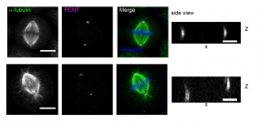
Figure 2: Regulation of mitotic spindle orientation by the actin-binding protein MISP. HeLa cells were transfected with Flag or Flag-MISP for 48 h and plated onto fibronectin-coated coverslips. Stainings for α-tubulin (green) and pericentrin (PCNT, magenta), co-stained with Hoechst (DNA, blue). Scale bars, 5 µm.
© dkfz.de
Mechanisms of drug resistance in cancer therapy
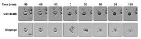
Spindle poisons are drugs that prevent the assembly (nocodazole, vincristine) or the disassembly (taxol or paclitaxel) of microtubules. They impair spindle function and chromosome segregation during mitosis by preventing normal microtubule dynamics. Treatment with spindle poisons therefore activates the spindle assembly checkpoint (SAC) leading to mitotic arrest. Those cells frequently undergo mitotic cell death. The microtubule poisons paclitaxel or vincristine are commonly used in chemotherapy in order to prevent proliferation of cancer cells by inducing cell death. Cells that are arrested in mitosis sometimes evade mitotic cell death. Instead these cells leave mitosis without completing a normal cell division and become tetraploid. This phenomenon is called mitotic slippage. During chemotherapy with spindle poisons cancer cells may evade mitotic cell death by performing mitotic slippage. This in turn leads to chemoresistance of cancer cells which continues to be a major impediment in medical oncology.
The goal of this project is to decipher the mechanism underlying chemoresistance (mitotic drug resistance) by analyzing the regulation of the F-box protein and tumor suppressor Fbxw7 during mitosis.
 more
more
Figure 3: Spindle poison treatment can lead to either apoptosis or mitotic slippage. Representative images from live-cell imaging are shown. 0 time point marks induction of mitotic cell death or mitotic slippage. Scale bars: 20 µm.
© dkfz.de
Our research activities are currently supported by the Deutsche Krebshilfe, the BMFT and the Deutsche José Carreras Leukämie-Stiftung e.V.