MRI Modern imaging modality without x-rays
Magnetic resonance imaging (MRI) is based on nuclear magnetic resonance (NMR) and allows imaging of the body without the application of x-rays. The MR scanner consists of a superconducting magnet and a high frequency (HF) or radiofrequency (RF) system, the latter operates similar to a radio transmitter or receiver. The magnetic field strength and radio waves used have no known harmful effects on the human body. The basic principle is the alignment of the spins of the atomic nuclei in the strong magnetic field of the scanner. The RF system stimulates the atomic nuclei by transmitting at the resonance frequency. The nuclei leave their stable alignment and begin to precess (rotate) like spinning tops. They dissipate the excess energy in the form of radio waves of the same frequency while slowly approaching the stable position again. The received frequencies can be encoded by the gradient system such that images can be reconstructed, which is usually done using a Fourier transform.
Considerations before performing an MRI exam
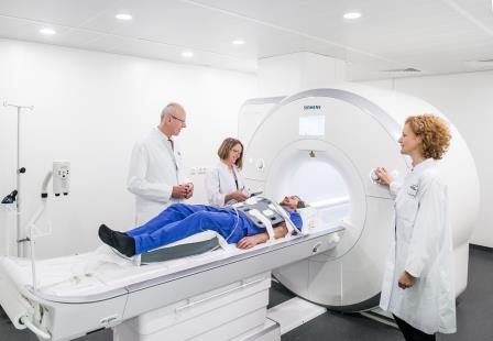
Aera 1.5 Tesla (Siemens)
- The magnetic field of the scanner is much stronger than the magnetic fields occurring in our regular daily experience. Metallic objects can be attracted by the magnetic field with high forces, potentially leading to injury. The RF energy can lead to heating of conductive materials (also non-ferromagnetic metals) and can disturb or destroy electronic circuits. Certain medical devices and implants (cardiac pacemakers, implanted cardioverters/defibrillators (ICD), electronic implants such as spinal stimulators, cochlear implants or similar, magnetically activated implants such as implanted insulin pumps or medication infusion pumps or similar, metal shrapnel or foreign bodies, non-MR compatible prostheses or implants) cannot be examined in the MR system.
- Metallic objects within the magnetic field can lead to injuries and imaging artifacts. All metallic or magnetic objects must be removed prior to the examination, including e.g. watches, glasses, keys, piercings, jewelry, hair pins and clips, purses and wallets, loose coins in pockets, credit cards (the magnetic information on the card will be erased by the MR field and the function of the card will be destroyed), metallic parts on clothing (belt, zipper, metallic buttons, bras), removable dentures, dental braces, hearing aids and acupuncture needles.
- Impaired renal function requires to be excluded before the administration of MR contrast agents. Appropriate recently performed blood tests should be obtained prior to the examination.
- The MR system requires lying on a table within a tunnel-shaped so called gantry. Some people with claustrophobia may not be able to tolerate lying in a confined space. In some instances a mild sedative may be prescribed. In these cases an accompanying person should be present and operating machinery such as cars will not be possible for a period of time, usually 24 hours. During this time please do not perform strenuous activities or engage in activities that require responsibility.
Considerations during an MR exam
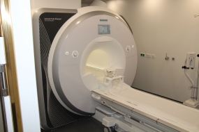
Prisma 3 Tesla MRT (Siemens)
© dkfz.de
The main MR magnet looks like a tube/tunnel with a 60-80 cm wide opening into which the table on which the patient is positioned can be introduced. During the examination you will usually hear loud noises, often resembling repeated knocking sounds. These are normal and are caused by electromagnetic switches (gradients) that are needed for imaging. The rate and volume of these noises varies during one and between examinations, depending on the body region examined and the protocol used. For comfort and hearing protection you will be offered ear plugs and/or ear phones. It is important that you remain as still as possible during the examination as individual images usually require several minutes and any motion will cause blurring or artifacts on the images that can impair the interpretation. For some examinations you may be given breathing instructions over the loudspeakers in the room or electrodes may be placed on your chest to obtain an electrocardiogram (ECG) during the scan to optimize the scan quality. On average the total examination time within the scanner is between 30 to 50 minutes, in some instances longer. During this time usually several hundred images are acquired. You have a call button in your hand and can signal to the MR operators at any time. Communication is possible through loudspeakers and microphones in the scanner.
Considerations after an MR examination
- If you received a sedative medication for the examination you will not be able to conduct any machinery including automobiles and you are not allowed to actively participate in traffic for 24 hours. During the same period you are not able to perform responsible activities. You are required to be accompanied by another person to the examination that will bring you home and remain with you overnight.
- If you are scheduled for two or more examinations at DKFZ on the same day, a small catheter will remain (usually in your arm) between the examinations.