Division of Radiology
Prof. Dr. med. Dipl.-Phys. Heinz-Peter Schlemmer
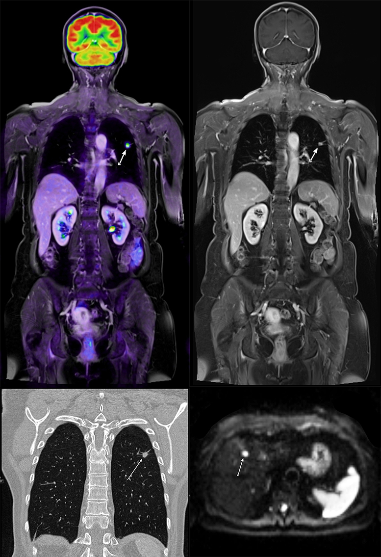
Multiparametric morphologic-functional imaging of lung cancer by CT, MR and PET/MR
© dkfz.de
Goal of the division of Radiology is to develop, validate and clinically implement imaging methods for improved cancer diagnostics and therapy and to transfer the technologies into the society and healthcare system. The division is rooted within the comprehensive research topic Imaging and Radiooncology with its strengths in medical physics, radiopharmacy, (radio)biology and bioinformatics, and complements the shared infrastructure with expertise to conduct clinical trials.
Strong multidisciplinar interactions close the gap between discovery, technological developments and clinical translation enabling both cross-fertilization of diverse research areas and clinical partners as well as backward translation from the clinics into further methodological advances. Our research on cross-sectional imaging technologies US, CT, MRI, PET/MRI including Artificial Intelligence (AI) can roughly be assigned to two categories:
(1) Research on new imaging methods to develop new prognostic/predictive imaging biomarkers for cancer detection, biological characterization, staging as well as individualized therapy planning and monitoring.
(2) Research in clinical trials by applying these innovations for exploring and visualizing cancer characteristics, for planning and monitoring response to novel therapies and for cancer screening.
We use the most recent cross-sectional imaging techniques including ultrasound, spectral CT, MR (1.5 T, 3.0 T, 7.0 T), simultaneous PET/MR and PET/CT. Scientific fields of application in oncology include, amongst others, prostate cancer, breast cancer, lung cancer, multiple myeloma and brain tumors. In close collaboration with the divisions of medical physics, medical informatics and radiation oncology tools for tumor diagnosis and quantification of individual therapy responses are developed and applied, to better assess changes in tissue composition during and after treatment and to perform improved quantification of therapy response. Within these collaborations further activities include computer-aided image analysis and computer assisted imaging applications for biopsy and treatment planning, e.g. for improving radiotherapy, or the detection and targeting of prostate cancer. Clinical studies are intertwined with patient care for which state-of-the-art oncologic imaging is provided in accordance with medical guidelines and regulatory requirements. Close cooperation exists in particular with the University Hospitals in Heidelberg and the DKFZ-Hector Cancer Institute in Mannheim. The DKFZ radiologists serve in multidisciplinary tumor board teams at the NCT Heidelberg, and the residents are enrolled in a cooperative residency program including the radiology departments of the University Hospital. Our division is involved in the German translational cancer research networks NCT and DKTK, the European network "Cancer Core Europe (CCE)", and of large-scale national BMBF- (German Federal Ministry of Education and Research) and international EU-funded projects on cancer imaging. Knowledge transfer into clinical practice and education is promoted, also in regions and countries with limited medical infrastructure and lower standards in Europe, Chile and Africa.
