B cells promote liver cancer with dangerous dual strategy
Inflammatory fatty liver disease (NASH, non alcoholic steatohepatitis ) and the resulting liver cancer are driven by autoaggressive T cells. Scientists from the German Cancer Research Center (DKFZ) now show what ist behind this destructive behavior. In both mice and humans with NASH, they found increased numbers of activated B cells in the gastrointestinal tract. The B cells promote the development of liver cancer with a dual strategy: via direct cell-cell contact, they activate autoaggressive T cells. In addition, the B cells produce IgA class antibodies that activate specific immune cells, thereby driving liver fibrosis. When the B cells are turned off, inflammation and fibrosis regress in mice and fewer and smaller liver tumors develop.
© Adobe Stock
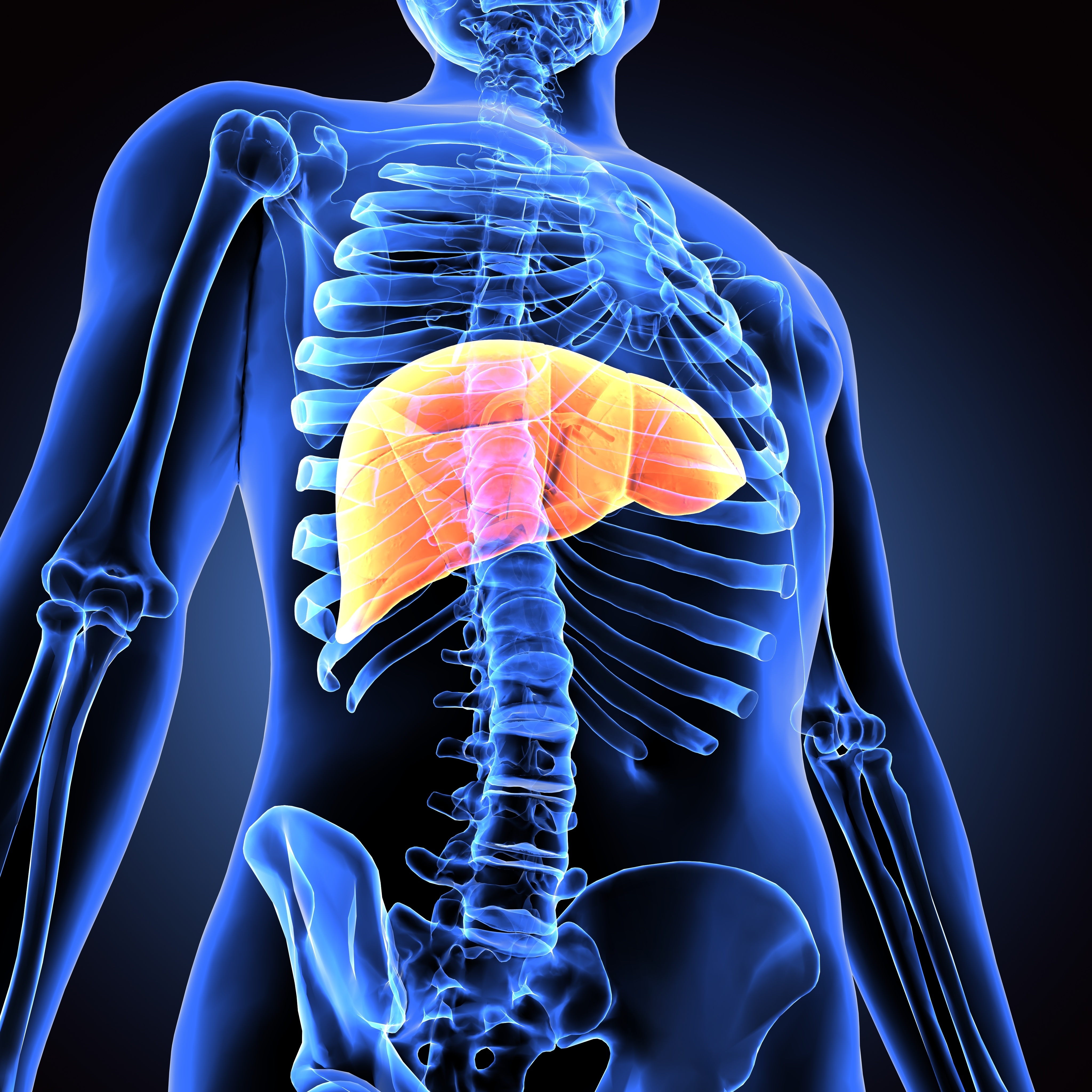
Liver cancer is the fourth leading cause of cancer death worldwide. The disease is driven by chronic inflammation triggered, for example, by viral infections or alcohol abuse. Very often, an unhealthy lifestyle is also the underlying cause: Too many calories, too little exercise and too high a body weight lead to a fatty liver. This, in turn, can result in non-alcoholic liver inflammation, also known as NASH, a veritable breeding ground for liver cancer.
"Worldwide, fatty liver and NASH are assuming pandemic proportions," says Mathias Heikenwälder of the German Cancer Research Center, a specialist in the links between chronic inflammation and liver cancer. Recently, a research team led by him found that NASH is driven by autoaggressive T cells that promote inflammatory tissue damage and even cancer development in the liver*.
What drives T cells to behave in this destructive manner? Several experimental findings suggested that B cells producing IgA class antibodies play a role in this process. Heikenwälder's team investigated this suspicion in a recent study in mice.
In mice fed a high-fat diet, inflammatory liver disease develops - much like in humans - and the animals often develop liver cell cancer. In contrast, mice that are genetically unable to produce B cells do not develop the disease under the same diet. In the livers of mice suffering from NASH, the researchers found a greatly increased number of activated B cells.
The B cells exert their disastrous influence on the liver in two ways: In the small intestine, they instigate T cells to behave autoaggressively via direct cell-cell contacts. The researchers were also able to reproduce this in the culture dish when they brought B cells from NASH mice together with CD8 T cells from a healthy animal, which were thereby activated to autoaggressive behavior.
In addition, the immunoglobulin A (IgA) produced by the B cells activates another group of immune cells, macrophages, which carry special IgA receptors on their surface. The activated macrophages aggravate fibrotic changes in the liver. If the B cells are switched off with a specific antibody in the NASH-afflicted animals, both the inflammation driven by the autoreactive T cells and the fibrosis regress.
Heikenwälder's team also examined tissue samples from people who had undergone surgery on the gastrointestinal tract to reduce weight ("bariatric surgery"). The findings strongly resembled those of mice suffering from NASH: Compared with healthy individuals, the tissue from NASH patients contained significantly more B cells, higher IgA levels and a higher number of activated macrophages.
"The results clearly show us that B cells as well as IgA are required to drive the pathological cascade in the development of liver cancer," Mathias Heikenwälder summarizes. "The good thing is that these results show us new ways to preventively interrupt this cancer-driving cascade: If we switch off the B cells with antibodies, the NASH symptoms regress and the animals develop fewer and smaller cancer foci. Fortunately, approved drugs already exist that suppress B-cell activation and that could possibly also stop NASH in humans and thus perhaps also liver cancer. However, there are no results from human studies on this yet."
Current publication:
Elena Kotsiliti, Valentina Leone, Svenja Schühle, Olivier Govaere, Hai Li, Monika Wolf ,Helena Horvatic, Sandra Bierwirth, Jana Hundertmark, Donato Inverso, Laimdota Zizmare, Avital Sarusi-Portuguez, Revant Gupta, Tracy O'Conor, Anastasios D. Giannou, Ahmad Mustafa Shiri, Maria Garcia Beccaria, Charlotte Rennert, Dominik Pfister, Rupert Öllinger, Iana Gadjalova, Pierluigi Ramadori, Mohammad Rahbari, Nuh Rahbari, Marc Healy Mirian Fernandez, Neda Yahoo, Jakob Janzen, Indrabahadur Singh, Chaofan Fan, Xinyuan Liu, Monika Rau, Martin Feuchtenberger, Eva Schwaneck, Sebastian J Wallace, Simon Cockell John Wilson-Kanamori, Prakash Ramachandran, Celia Kho, Timothy J Kendall, Anne-Laure Leblond, Selina J. Keppler, Piotr Bielecki, Katja Steiger, Maike Hofmann, Karsten Rippe, Horst Zitzlesberger, Achim Weber, Nisar Malek, Tom Luedde, Mihael Vucur, Hellmut G.Augustin, Richard Flavell, Oren Parnas, Roland Rad, Olivier Pabst, Neil C. Henderson, Samuel Huber, Andrew Macpherson, Percy Knolle, Manfred Claasen, Andreas Geier, Christoph Trautwein, Kristian Unger, Eran Elinav, Ari Waisman, Zeinab Abdullah, Dirk Haller, Frank Tacke, Quentin M. Anstee, and Heikenwälder Mathias: Gastrointestinal B-cells license metabolic T-cell activation 1 in NASH microbiota/antigen independently and contribute to fibrosis by IgA-FcR signaling.
Journal of Hepatology 2023, DOI: https://doi.org/10.1016/j.jhep.2023.04.037
* Dudek M. et al.: Auto-aggressive CXCR6+ CD8 T cells cause liver immune pathology in NASH.
Nature 2021, DOI: 10.1038/s41586-021-03233-8
Pfister D. et al. NASH limits anti-tumour surveillance in immunotherapy-treated HCC.
Nature 2021, DOI 10.1038/s41586-021-03362-0
With more than 3,000 employees, the German Cancer Research Center (Deutsches Krebsforschungszentrum, DKFZ) is Germanys largest biomedical research institute. DKFZ scientists identify cancer risk factors, investigate how cancer progresses and develop new cancer prevention strategies. They are also developing new methods to diagnose tumors more precisely and treat cancer patients more successfully. The DKFZ's Cancer Information Service (KID) provides patients, interested citizens and experts with individual answers to questions relating to cancer.
To transfer promising approaches from cancer research to the clinic and thus improve the prognosis of cancer patients, the DKFZ cooperates with excellent research institutions and university hospitals throughout Germany:
The DKFZ is 90 percent financed by the Federal Ministry of Education and Research and 10 percent by the state of Baden-Württemberg. The DKFZ is a member of the Helmholtz Association of German Research Centers.