PSMA-binding agents: versatile against prostate cancer
PSMA-binding agents specifically dock onto prostate cancer cells. Coupled to diagnostic or therapeutic radionuclides, they can improve the diagnosis and treatment of prostate cancer. Scientists from the DKTK partner site in Freiburg, together with scientists from the Max Planck Institute for Medical Research, have now used STED microscopy to investigate for the first time how these substances are taken up by the cell and distributed intracellularly. In addition, a first clinical application showed that hybrid PSMA-binding agents containing both a diagnostic radionuclide and a fluorescent dye are suitable for visualizing prostate cancer both before and during surgery.
In the DKTK, the German Cancer Research Center (DKFZ) in Heidelberg, as the core center, joins forces in the long term with university partner sites in Germany that have a special reputation for oncology.
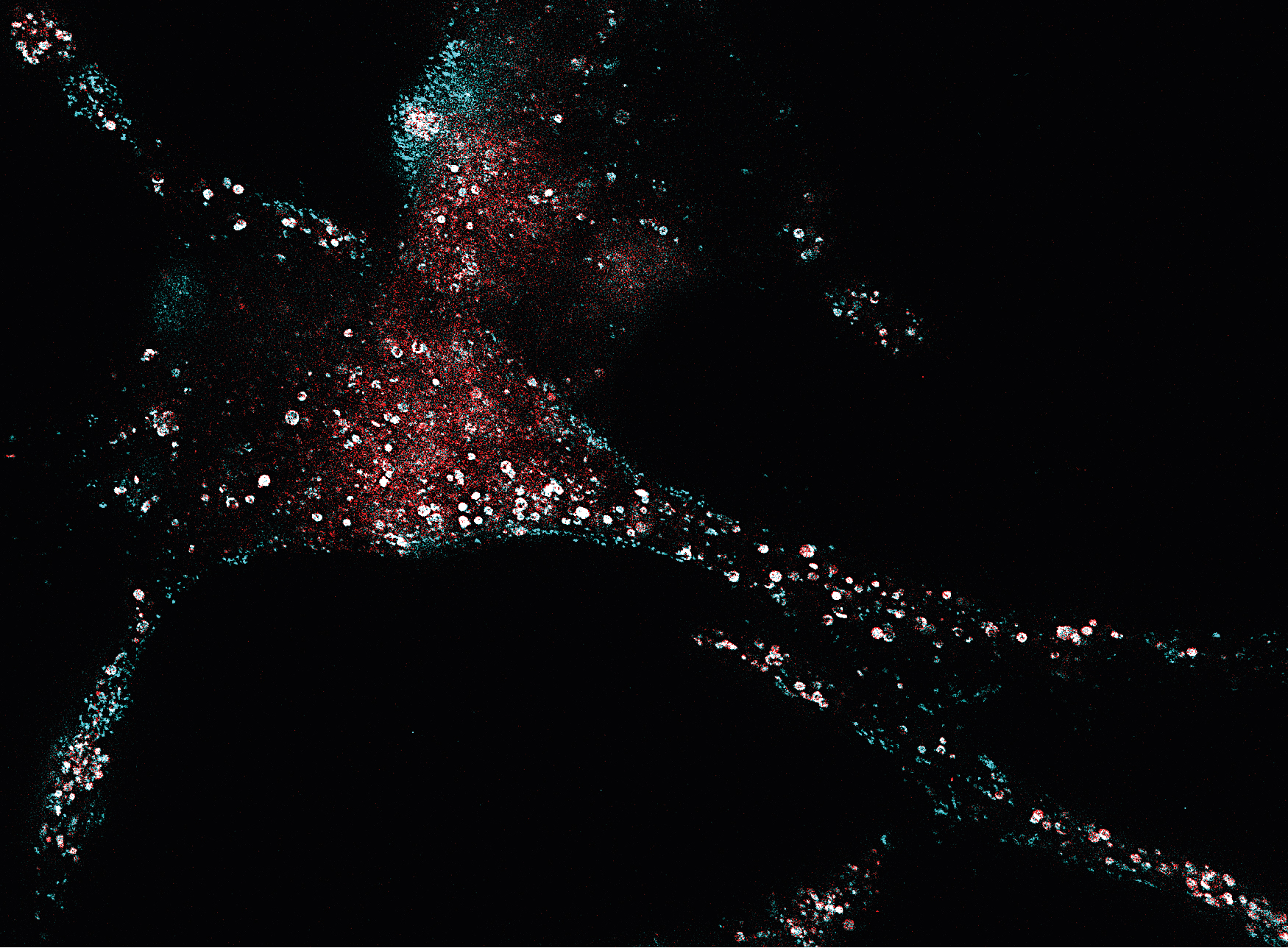
STED microscopy can be used to study the distribution and accumulation of PSMA-binding agents (red) in prostate cancer cells. In comparison: The distribution of PSMA (cyan).
© Ann-Christin Eder, DKTK and Jessica Matthias, MPI
PSMA, the prostate-specific membrane antigen, is present in small amounts on the surface of healthy prostate cells, but is much more abundant on prostate cancer cells. The protein is hardly found in the rest of the body. PSMA is therefore an ideal marker for the diagnosis of prostate cancer and at the same time a suitable target structure for specific therapies against the disease.
In recent years, substances have been developed at the German Cancer Research Center (Deutsches Krebsforschungszentrum, DKFZ) and Heidelberg University Hospital that specifically dock to PSMA and can be labeled with various radioactive substances known as radionuclides. "These radionuclide-coupled agents can be used to irradiate cancer cells from the inside," explains Ann Christin Eder, a scientist at the DKTK partner site in Freiburg at the University Medical Center Freiburg. "For this to work, the PSMA-binding agents must first be taken up into the cancer cell and remain there for as long as possible."
This process has been very little studied so far. In collaboration with Jessica Matthias and other scientists from the Max Planck Institute for Medical Research, Eder and her team have now used a method which allows precise insights into living cells in a nanometer scale to precisely analyze the distribution of the agents in the cell 1): STED microscopy STED microscopy, for which Stefan Hell, then of DKFZ and the Max Planck Society, was awarded the Nobel Prize in 2014.
For the current study, the research team led by Ann-Christin Eder at the DKFZ in Heidelberg and in their laboratory in Freiburg used so-called hybrid PSMA-binding molecules that can be coupled simultaneously with two different markers: In addition to a diagnostic radioactive label, they also bind a fluorescent dye, which also enables their use in STED microscopy.
The researchers' most important finding was that the PSMA-binding agents remained in the prostate cancer cells for a long time and accumulated there more and more over time. The molecules distributed homogeneously in the cytoplasm which may be advantageous for their therapeutic application..
The hybrid PSMA-binding agents, which consist of both radioactive and fluorescent markers, are considered promising tools to improve prostate cancer diagnosis and therapy. Through their radioactive labeling, they serve as tracers through which the tumor and its metastases can be localized using a combination of positron emission tomography (PET) and computed tomography (CT). This non-invasive imaging can be used to plan surgery and radiation therapy.
During surgery, fluorescent dye coupled to the pharmacon then helps surgeons distinguish between malignant and healthy tissue so they can precisely remove the tumor. This approach, which visualizes prostate cancer before and during surgery, was recently successfully tested for the first time with the hybrid agent PSMA-914 developed by Eder and her team in a patient with aggressive prostate cancer at the Clinics of Nuclear Medicine and Urology, University Hospital Freiburg2). PSMA-914 contains 68gallium as a diagnostic radionuclide, as well as a fluorescent dye.
"This first clinical application proves to us the potential of hybrid agents," explains Ann Christin Eder. "Future studies will now show whether PSMA-914 can also lead to improved therapeutic outcomes."
1)Jessica Matthias, Johann Engelhardt, Martin Schäfer, Ulrike Bauder-Wüst, Philipp T. Meyer, Uwe Haberkorn, Matthias Eder, Klaus Kopka, Stefan W. Hell, Ann-Christin Eder: Cytoplasmic localization of prostate-specific membrane antigen inhibitors may confer advantages for targeted cancer therapies
Cancer Research 2021, DOI: 10.1158/0008-5472.CAN-20-1624
2)Ann-Christin Eder, Mohamed A. Omrane, Sven Stadlbauer, Mareike Roscher, Wael Y. Khoder,Christian Gratzke, Klaus Kopka, Matthias Eder & Philipp T. Meyer, Cordula A. Jilg, Juri Ruf: The PSMA-11-derived hybrid molecule PSMA-914 specifically identifies prostate cancer by preoperative PET/CT and intraoperative fluorescence imaging
European Journal of Nuclear Medicine and Molecular Imaging 2021,
DOI: 10.1007/s00259-020-05184-0
A picture is available for download:
www.dkfz.de/de/presse/pressemitteilungen/2021/bilder/PSMA-bindende-Wirkstoffe.jpg
Picture Caption: STED microscopy can be used to study the distribution and accumulation of PSMA-binding agents (red) in prostate cancer cells. In comparison: The distribution of PSMA (cyan).
Note on use of images related to press releases
Use is free of charge. The German Cancer Research Center (Deutsches Krebsforschungszentrum, DKFZ) permits one-time use in the context of reporting about the topic covered in the press release. Images have to be cited as follows: "Source: Ann-Christin Eder, DKTK and Jessica Matthias, MPI".
Distribution of images to third parties is not permitted unless prior consent has been obtained from DKFZ's Press Office (phone: ++49-(0)6221 42 2854, E-mail: presse@dkfz.de). Any commercial use is prohibited.
With more than 3,000 employees, the German Cancer Research Center (Deutsches Krebsforschungszentrum, DKFZ) is Germanys largest biomedical research institute. DKFZ scientists identify cancer risk factors, investigate how cancer progresses and develop new cancer prevention strategies. They are also developing new methods to diagnose tumors more precisely and treat cancer patients more successfully. The DKFZ's Cancer Information Service (KID) provides patients, interested citizens and experts with individual answers to questions relating to cancer.
To transfer promising approaches from cancer research to the clinic and thus improve the prognosis of cancer patients, the DKFZ cooperates with excellent research institutions and university hospitals throughout Germany:
The DKFZ is 90 percent financed by the Federal Ministry of Education and Research and 10 percent by the state of Baden-Württemberg. The DKFZ is a member of the Helmholtz Association of German Research Centers.