3D atlas of the bone marrow - in single cell resolution
Stem cells located in the bone marrow generate and control the production of blood and immune cells. Researchers from EMBL, DKFZ and HI-STEM have now jointly developed new methods to reveal the three-dimensional organization of the bone marrow at the single cell level. Using this approach the teams have identified previously unknown cell types that create specific local environments required for blood generation from stem cells. The study, published in Nature Cell Biology, reveals an unexpected complexity of the bone marrow and its microdomains at an unprecedented resolution and provides a novel scientific basis to study blood diseases such as leukemias.
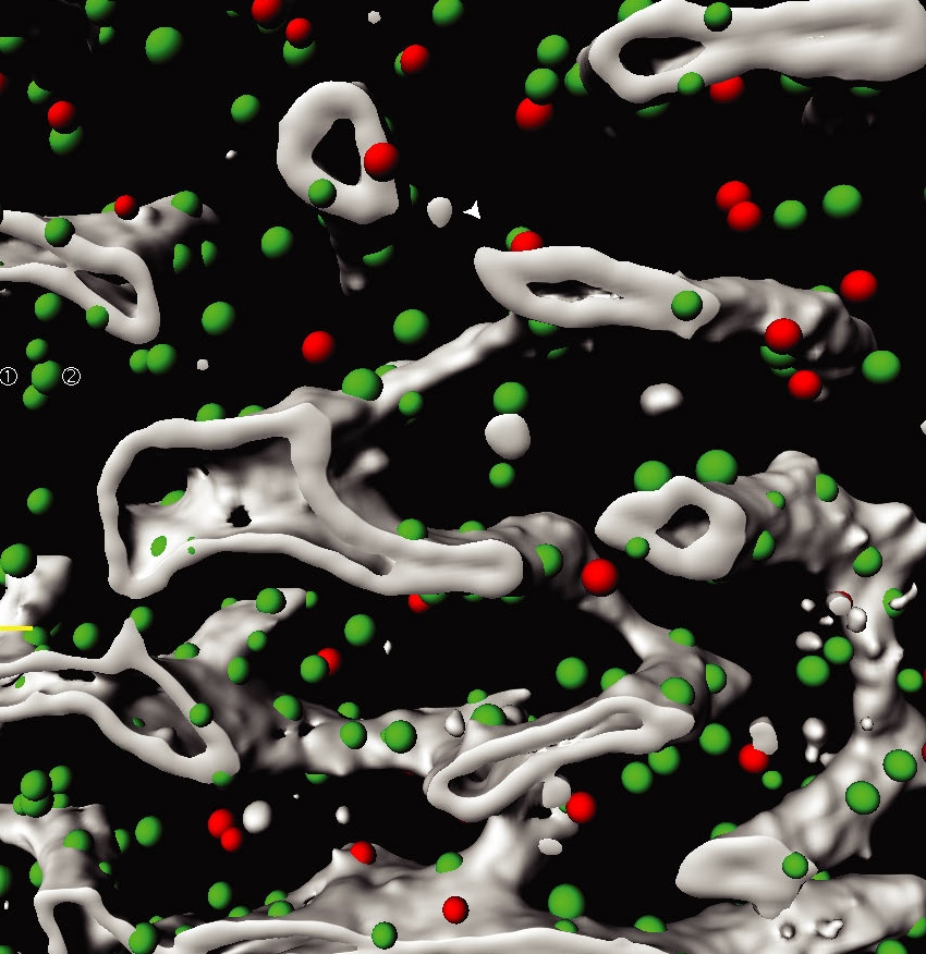
Three dimensional segmentation of a bone marrow region. Different niche cells (green and red dots) and blood vessels (grey) are highlighted.
© DKFZ, EMBL and University Hospital Zurich
In the published study researchers from European Molecular Biology Laboratory (EMBL), the German Cancer Research Center (DKFZ) and the Heidelberg Institute for Stem Cell Technology and Experimental Medicine* (HI-STEM gGmbH) present new methods permitting the characterisation of complex organs. The team focused their research on the murine bone marrow, as it harbours blood stem cells that are responsible for life-long blood production. Because of the ability to influence stem cells and to sustain blood production, there is a growing interest in exploiting the bone marrow environment, also called niche, as a target for novel leukaemia treatments. "So far, very little was known about how different cells are organised within the bone marrow and how they interact to maintain blood stem cells," explains Chiara Baccin, post-doc in the Steinmetz Group at EMBL. "Our approach unveils the cellular composition, the three-dimensional organisation and the intercellular communication in the bone marrow, a tissue that has thus far been difficult to study using conventional methods," further explains Jude Al-Sabah, PhD student in the Haas Group at HI-STEM and DKFZ.
In order to understand which cells can be found in the bone marrow, where they are localised and how they might impact on stem cells, the researchers combined single-cell and spatial transcriptomics with novel computational methods. By analysing the RNA content of individual bone marrow cells, the team identified 32 different cell types, including extremely rare and previously unknown cell types. "We believe that these rare 'niche cells' establish unique environments in the bone marrow that are required for stem cell function and production of new blood and immune cells," explains Simon Haas, group leader at the DKFZ and HI-STEM, and one of the initiators of the study.
Using novel computational methods, the researchers were not only able to determine the organisation of the different cell types in the bone marrow in 3D, but could also predict their cellular interactions and communication. "It's the first evidence that spatial interactions in a tissue can be deduced computationally on the basis of genomic data," explains Lars Velten, staff scientist in the Steinmetz Group.
"Our dataset is publicly accessible to any laboratory in the world and it could be instrumental in refining in vivo studies," says Lars Steinmetz, group leader and director of the Life Science Alliance at EMBL Heidelberg. The data, which is now already used by different teams all over the world, is accessible via a user-friendly web app.
The developed methods can in principle be used to analyse the 3D organisation of any organ at the single cell level. "Our approach is widely applicable and could also be used to study the complex pathology of human diseases such as anemia or leukemia" highlights Andreas Trumpp, managing director of HI-STEM and division head at DKFZ.
*The Heidelberg Institute for Stem Cell Research and Experimental Medicine (HI-STEM) gGmbH was founded in 2008 as a public-private partnership between the DKFZ and the Dietmar Hopp Foundation
Chiara Baccin, Jude Al-Sabah, Lars Velten, Patrick M. Helbling, Florian Grünschläger, Pablo Hernández-Malmierca, César Nombela-Arrieta, Lars M. Steinmetz, Andreas Trumpp, and Simon Haas: Combined single-cell and spatial transcriptomics reveal the molecular, cellular and spatial bone marrow niche organization.
Nature Cell Biology 2019, https://doi.org/10.1038/s41556-019-0439-6
A picture is available for Download:
www.dkfz.de/de/presse/pressemitteilungen/2019/bilder/Illustration-BM.jpg
Picture Caption: Three dimensional segmentation of a bone marrow region. Different niche cells (green and red dots) and blood vessels (grey) are highlighted.
Note on use of images related to press releases
Use is free of charge. The German Cancer Research Center (Deutsches Krebsforschungszentrum, DKFZ) permits one-time use in the context of reporting about the topic covered in the press release. Images have to be cited as follows: "Source: DKFZ, EMBL and University Hospital Zurich".
Distribution of images to third parties is not permitted unless prior consent has been obtained from DKFZ's Press Office (phone: ++49-(0)6221 42 2854, E-mail: presse@dkfz.de). Any commercial use is prohibited.
With more than 3,000 employees, the German Cancer Research Center (Deutsches Krebsforschungszentrum, DKFZ) is Germanys largest biomedical research institute. DKFZ scientists identify cancer risk factors, investigate how cancer progresses and develop new cancer prevention strategies. They are also developing new methods to diagnose tumors more precisely and treat cancer patients more successfully. The DKFZ's Cancer Information Service (KID) provides patients, interested citizens and experts with individual answers to questions relating to cancer.
To transfer promising approaches from cancer research to the clinic and thus improve the prognosis of cancer patients, the DKFZ cooperates with excellent research institutions and university hospitals throughout Germany:
The DKFZ is 90 percent financed by the Federal Ministry of Education and Research and 10 percent by the state of Baden-Württemberg. The DKFZ is a member of the Helmholtz Association of German Research Centers.