Endothelial cells in the driver's seat
Endothelial cells, the inner lining of blood vessels, do not just respond passively to exogenous stimuli. Instead, they themselves control organ function in a very active manner. Scientists from the German Cancer Research Center and Heidelberg University have now discovered a complex growth regulatory mechanism through which endothelial cells control liver regeneration after damage or partial surgical removal.
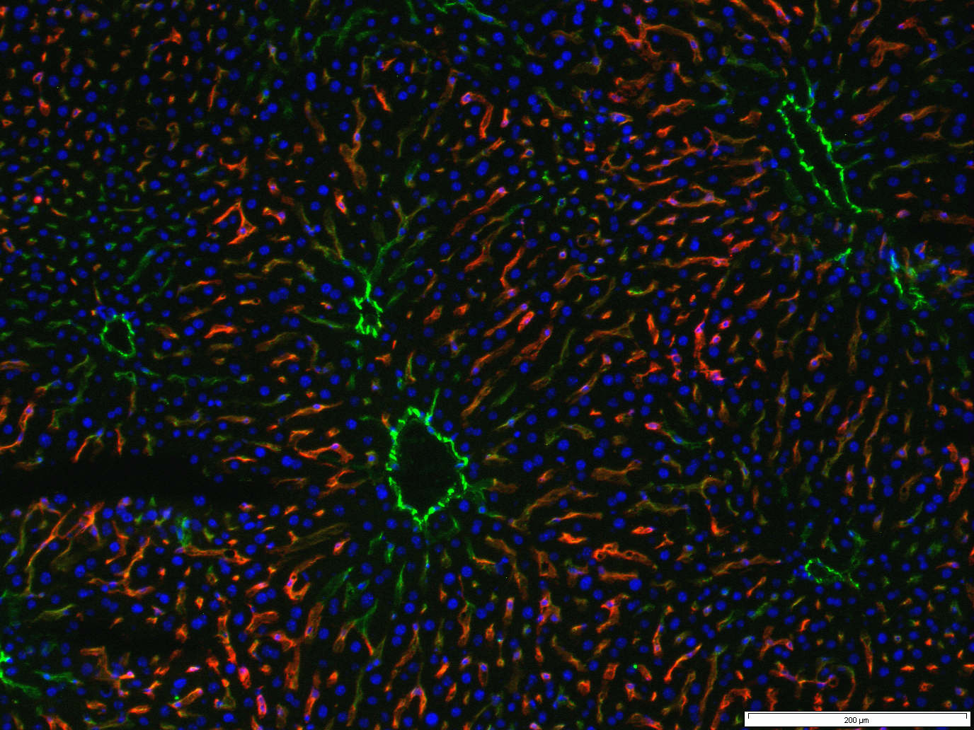
Histologic image showing the lobular microarchitecture of the liver. Hepatocytes (blue nuclei) are in close contact with sinusoidal endothelial cells (red; Lyve-1 staining). Larger liver vessels including the central vein of liver lobes as well as portal arteries and veins express the differentiation marker CD31 (green).
© dkfz.de
A dense network of arteries, capillaries and veins leaves every cell of the body within less than one tenth of a millimeter of the nearest blood vessel. A single layer of cells, called endothelial cells, covers the inner lining of all blood vessels. Vascular dysfunction is critically involved in some of the most devastating human diseases, including cardiac ischemia, atherosclerosis, stroke and the growth of tumors. In fact, endothelial cells are directly or indirectly involved in more than two thirds of all human causes of death. The better molecular and mechanistic understanding of the complexity of endothelial cell function consequently holds great promise to lead to the development of novel therapies.
Endothelial cells are widely considered as a rather passive, barrier-forming cell population that at best responds to exogenous stimuli. Recent work is casting a very different view of endothelial cells: Instead of just serving as passive responder to exogenous stimuli, endothelial cells themselves control surrounding cells quite actively.
A team of scientists from the German Cancer Research Center and the Medical Faculty Mannheim of Heidelberg University headed by Professor Hellmut Augustin has systematically studied the role of the vascular endothelium as a regulator and gatekeeper of liver regeneration. The findings of this study have now been published in the scientific magazine Science.
The scientists surgically removed in healthy mice two third of the liver. This loss of metabolic function induces a massive proliferation of the remaining liver cells (hepatocytes). As a result, normal liver size is restored within few days. To study the orchestrating role of the vascular endothelium, the changes in gene expression of isolated liver endothelial cells one day after surgery were analyzed. Of the identified up- and downregulated genes, Angiopoietin-2 (Ang2) was identified as a key regulator of liver regeneration.
Ang2 is produced by endothelial cells and controls the maturation and quiescence of blood vessels. Upon partial hepatectomy, Ang2 production in endothelial cells is rapidly downregulated. As a consequence, the expression of genes that are regulated by Ang2 is also altered. Among these is TGFß, which acts as a well-established inhibitor of hepatocyte proliferation. As such, the work revealed that liver endothelial cells control hepatocyte proliferation during liver regeneration by blocking an inhibitor pathway that acts as an endogenous brake.
But that’s only part of the story: Additional experiments revealed that endothelial cell produced Ang2 does not just control hepatocyte proliferation, but also supports the subsequent regeneration of the associated connective tissue. “Endothelial cells control liver regeneration as a dynamic rheostat” explains Dr. Junhao Hu, first authors of the study, the groundbreaking character of the discoveries. “By dynamically regulating Ang2 production, endothelial cells first control hepatocyte proliferation and later their own regeneration”. Endothelial cells do so, by again upregulating the expression of Ang2 following the hepatocyte regenerative phase. As a result, Ang2 controls endothelial cells expression of VEGF receptor-2. This in turn allows them to grow new blood vessels by responding to hepatocyte-produced angiogenesis-inducing VEGF growth factor.
The study is a prototypic example demonstrating how the translation of basic science findings to the analysis of the molecular mechanisms of diseases may yield surprising and unexpected discoveries. “We are only beginning to understand the full complexity of endothelial cell functions” explains Professor Augustin. “The newly emerging concepts of ‘angiocrine signaling’ - the notion that endothelial cells are active drivers and not just passive responders - will transform our conceptual understanding of vascular function and may in the long run pave the way towards the development of novel therapeutic strategies aimed at interfering with vessel and organ diseases.”
Hu J, Srivastava K, Wieland M, Runge A, Mogler C, Besemfelder E, Terhardt D, Vogel MJ, Cao L, Korn C, Bartels S, Thomas M, Augustin HG: Endothelial cell-derived Angiopoietin-2 controls liver regeneration as a spatiotemporal rheostat. Science, 2014, DOI: 10.1126/science.1244880.
A picture for this press release is available at:
http://www.dkfz.de/de/presse/pressemitteilungen/2014/bilder/10xtriple.jpg
Caption: Histologic image showing the lobular microarchitecture of the liver. Hepatocytes (blue nuclei) are in close contact with sinusoidal endothelial cells (red; Lyve-1 staining). Larger liver vessels including the central vein of liver lobes as well as portal arteries and veins express the differentiation marker CD31 (green).
Picture source: Dr. Junhao Hu/DKFZ
With more than 3,000 employees, the German Cancer Research Center (Deutsches Krebsforschungszentrum, DKFZ) is Germanys largest biomedical research institute. DKFZ scientists identify cancer risk factors, investigate how cancer progresses and develop new cancer prevention strategies. They are also developing new methods to diagnose tumors more precisely and treat cancer patients more successfully. The DKFZ's Cancer Information Service (KID) provides patients, interested citizens and experts with individual answers to questions relating to cancer.
To transfer promising approaches from cancer research to the clinic and thus improve the prognosis of cancer patients, the DKFZ cooperates with excellent research institutions and university hospitals throughout Germany:
The DKFZ is 90 percent financed by the Federal Ministry of Education and Research and 10 percent by the state of Baden-Württemberg. The DKFZ is a member of the Helmholtz Association of German Research Centers.