Functional
Genome Analysis (B070)
Deutsches
Krebsforschungszentrum,
Im Neuenheimer Feld 580
D-69120
Heidelberg,
Germany. |
 |
.
.
|
Archive |
Proteomics
- Antibody
Microarray Technology |
|
FINISHED PROJECT:
Utilisation of
antibody microarrays for the selection of specific and
informative antibodies from library binders of unknown quality
Many
diagnostic and therapeutic concepts require
antibodies of high specificity. Recombinant binder libraries and
related
selection approaches allow the isolation of antibodies against almost
every target of interest. Nevertheless, it cannot be guaranteed that
selected
antibodies perform well and interact specifically enough with analytes
unless
an elaborate characterisation is performed. Here, we present an
approach to
shorten this process by combining the selection of suitable antibodies
with the
identification of informative target molecules by means of antibody
microarrays, thereby reducing the effort of antibody characterisation
by
concentrating on relevant molecules.
..
In a pilot
scheme, a library of 456 single-chain
variable fragment (scFv) binders to 134 antigens was used. They were
arranged
in a microarray format and incubated with the protein content of
clinical
tissue samples isolated from pancreatic ductal adenocarcinoma and
healthy
pancreas, as well as recurrent and non-recurrent bladder tumours. We
observed
significant variation in the expression of the E3 ubiquitin-protein
ligase
(CHFR) as well as the glutamate receptor interacting protein 2 (GRIP2),
for
example, always with more than one of the scFvs binding to these
targets. Only
the relevant antibodies were then characterised further on antigen
microarrays
and by surface plasmon resonance experiments so as to select the most
specific
and highest affinity antibodies. These binders were in turn used to
confirm a
microarray result by immunohistochemistry analysis.
Kibat et
al. (2016) New
Biotechnol. 33,
574-581. 
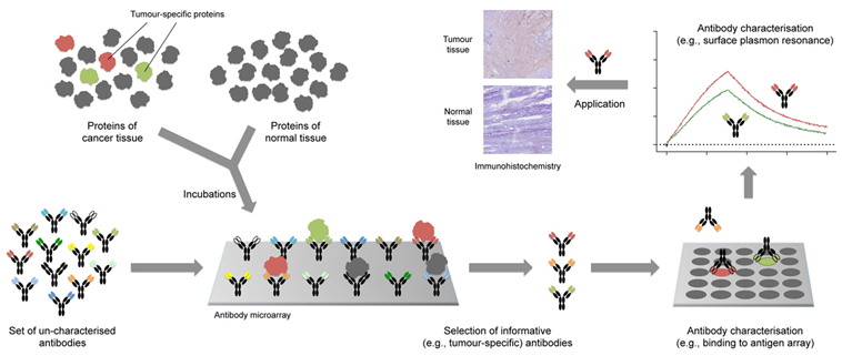
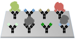
Scheme of the
selection and characterisation
process for a highly effective isolation of both specific and
informative antibodies
(or other binders) from a library of uncharacterised molecules. |
|
|
FINISHED PROJECT:
Affinomics: Proteome binders for
characterisation of human proteome function; generation, validation,
application
|


|
The Affinomics
programme aims
to leverage existing efforts in Europe to generate
large-scale resources of validated protein-binding
molecules (binders) as affinity reagents for characterisation of the
human
proteome and to apply them in comprehensive structural and functional
analyses
of protein expression, interactions and complexes. The project was
preceded by the ProteomeBinders and AffinityProteome consortia.
..
Proteome
targets will be
focused on five categories of inter-related human proteins involved in
signal
transduction, cell regulation and cancer, namely protein kinases, SH2
domain-containing proteins, protein tyrosine phosphatases, proteins
somatically
mutated in cancers and candidate cancer biomarkers. Binders to about
1000
protein targets will be made over the course of the programme.
..
A high
throughput, coordinated production pipeline for antigens and binders
will be
established. Target antigens will be expressed in three forms, as
folded
full-length proteins or domains, as large peptide fragments (PrESTs)
based on
low homology to other human proteins and as small peptides, in some
cases
phosphorylated. Binder types to be generated include affinity-purified
polyclonal antibodies, monoclonal antibodies, recombinant antibody
fragments
and non-immunoglobulin scaffolds.
..
An important
aspect will be the development of
highly efficient next generation recombinant selection methods, based
on phage,
cell and ribosome display, capable of producing high quality binders at
greater
throughput and lower cost than hitherto. Systems and procedures for
thorough
binder validation and quality control will be established. The affinity
reagents will be applied in advanced innovative and sensitive
technologies for
specific detection of target proteins and interacting protein complexes
in
cells, tissues and fluids, for improved understanding of protein
function and
new classes of diagnostic assays.
..
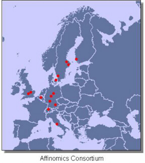
For more
detailed information, click on the map of the consortium.
|
Sequence
validation of binders
..
Antibodies and other binder
types are crucial for any proteome analysis. Although many tens of
thousands of antibodies are available from commercial sources and academic institutions (see antibodypedia,
for example),
quality and
reproducibility of analyses performed with these molecules vary
substantially. Antibodies exhibit
huge differences in specificity and affinity, including binders that target the same protein. Worse, even binders
that are supposedly from the same source showed significant lot-to-lot
variation.
..
One reason for
variation could be the
actual assay; an antibody
perfoming very well on Western blots may not meet the quality standards
of pull-down experiments or microarray analyses, and vice versa. Another point, however,
that is contributing significantly to low reproducibility is a lack of
commonly accepted performance parameters and tests for their definition
This is made worse by the fact that currently most binders are
ill-described. Consequently, one cannot be sure, if a binder is exactly
the molecule that was used in assays before.
..
Initiated by
Andrew Bradbury, 101 scientists of the Affinomics consortium and beyond
have had some thoughts about the matter. A simple solution to the
problem of how to make sure that everybody is really using the very
same
binder molecules in their experiments would be a sequence verification
of each binder. While relatively easily achievable for recombinant
binders and monoclonal antibodies, it is more difficult to establish
for polyclonal binders, for example. A more important obstacle for an
implementation of such
a scheme, however, could be the reluctance of antibody producers to
share the sequences of their molecules, since this would make available
their good binders to the entire scientific (and commercial) community
for free.
Bradbury et
al. (2015) Nature 518,
27-29.  |
FINISHED PROJECT:
Protein
profiling of gastric cancer and neighbouring control tissues
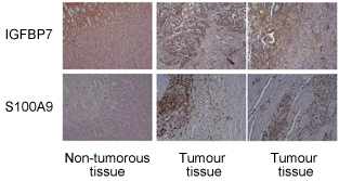
Protein
profiling was performed on gastric cancer tissue samples. Sixteen pairs
of
postoperative gastric adenocarcinomas and adjacent non-cancerous
control
tissues were analysed on microarrays that contain 813 antibodies
targeting 724
proteins. Only 17 proteins were found to be differentially regulated,
much fewer
molecules than the number identified when comparing tumour to healthy
control
tissues. Insulin-like growth factor-binding protein 7 (IGFBP7), S100
calcium
binding protein A9 (S100A9), interleukin-10 (IL-10) and mucin 6 (MUC6)
exhibited the most profound variations. For an evaluation of the
proteins’
capacity for discriminating gastric cancer, a Receiver Operating
Characteristic
curve analysis was performed. For confirmation, immunohistological
analyses
were done on samples prepared from another cohort of patients with
gastric
cancer.
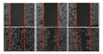
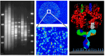
Figure legend:
Typical results of immunohistochemical analyses. Identified marker
molecules were validated by immunohistochemistry on an independent set
of tumour tissues and stomach samples from donors who had no cancer. Dark brown
colour is
indicative of the presence of the respective proteins.
Sill et
al. (2016) Microarrays 5,
19. |
|
|
..
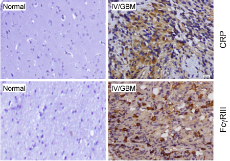
| Immunostaining of CRP and
its receptor FcγRIII was
performed in normal brain tissue (n= 17) and GBM tumor tissue sections
(n= 46);
representative images are shown. Both proteins were present in
microglial cells. |
|
|
|
FINISHED PROJECT:
Definition of a
serum marker panel for glioblastoma discrimination and identification
of interleukin 1β in the microglial
secretome as a novel mediator of endothelial cell survival induced by C-reactive
protein

Glioblastoma
(GBM) is the most common malignant
adult primary brain tumour. We profiled 724 cancer-associated proteins
in sera
of healthy individuals (n = 27) and GBM (n = 28) using an antibody
microarray.
While 69 proteins exhibited differential abundance in GBM sera, a
three-marker
panel (LYAM1, BHE40 and CRP) could discriminate GBM sera from that of
healthy
donors with an accuracy of 89.7% and p = 0.0001.
The high abundance of C-reactive protein (CRP) in
GBM sera was confirmed in 264 independent samples. High levels of CRP
protein
were seen in GBM but without a change in transcript levels suggesting a
non-tumoral origin. Glioma-secreted Interleukin 6 (IL6) was found to
induce
hepatocytes to secrete CRP, involving the JAK-STAT pathway. The culture
supernatant from CRP-treated microglial cells induced endothelial cell
survival
under nutrient-deprivation condition involving the CRP-FcγRIII
signalling
cascade. Transcript profiling of CRP-treated microglial cells
identified
Interleukin 1β (IL1β) present in the microglial secretome as the key
mediator
of CRP-induced endothelial cell survival. IL1β neutralization by
antibody-binding or siRNA-mediated silencing in microglial cells
reduced the
ability of the supernatant from CRP-treated microglial cells to induce
endothelial cell survival. Our study identifies a serum based
three-marker
panel for GBM diagnosis and provides leads for developing targeted
therapies.
.
Nijaguna et
al. (2015) J. Proteomics 128, 251-261. |
FINISHED PROJECT:
Prediction of
recurrence of non muscle-invasive bladder cancer
About
70% of newly diagnosed cases of bladder cancer are low-stage,
low-grade, non
muscle-invasive. Standard treatment is
transurethral resection. About 60% of the tumours will recur,
however,
and in part progress to become invasive. Therefore, surveillance
cystoscopy is
performed after resection. In the USA
and Europe alone,
about 54,000 new
patients per year undergo repeated cystoscopies over several years, who do not experience recurrence. Analysing
in a pilot study
resected tumours from patients with and without local recurrence after
a period
of five years, we identified 255 proteins with significantly
differential
abundance. Most are involved in the regulation and execution of
apoptosis and
cell proliferation. A multivariate classifier was constructed based on
20
proteins. It facilitates the prediction of recurrence with a
sensitivity of 80%
and a specificity of 100%. As a measure of overall accuracy, the area
under the
curve (AUC) value was found to be 91%. After
validation, such a signature could help to
adjust the treatment scheme and the rigidity of surveillance. This
could
dramatically reduce surveillance cost and simultaneously improve the
patients’
outcome substantially.
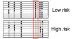
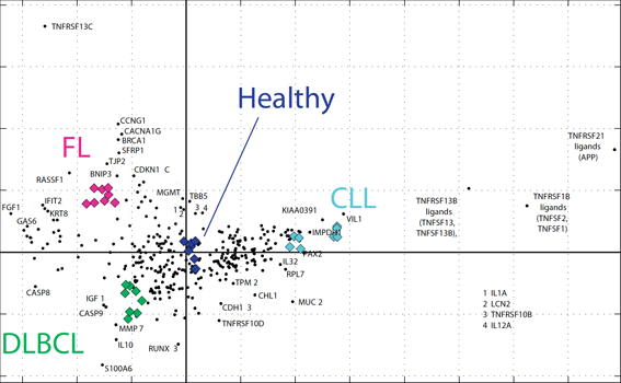
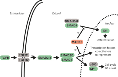
Figure legend: Strong
repression of the TGF-beta signalling
pathway in recurrent cancer. Affected proteins are
labelled in green or red if their expression was lower or higher,
respectively,
in recurrent rather than non-recurrent cancer.
Srinivasan et
al. (2014) Proteomics 14, 1333. |
|
|
FINISHED PROJECT:
Automated,
real-time and multiplex analysis of biomolecular interactions (ARTAMIS)
...

The goal of
the project was the
development of a real-time fluorescence detection system of
protein-ligand
interactions. The systems allows the effective combination of
different
molecular biology procedures with ultra-sensitive, multiplexed
detection, the
final objective being a real-time analysis of biomolecular interactions
for
virtually any kind of biomolecules under various conditions.
..
In recent
years, the detailed
study of biomolecular interactions has become a very important and
growing
element of biomedical research. This increased interest has been driven
by the appreciation
that a deep understanding of protein interactions is fundamental to
both the
study of disease and the elucidation of the action of small molecule
drugs or
biopharmaceuticals. Protein-based pharmaceuticals are currently
enjoying great
commercial success and are accounting for a growing proportion of the
pharmaceutical industry’s development portfolio.
..
Although
some detection schemes
and relevant equipment have been developed for the measurement of
biomolecular
interactions, a lot of progress remains to be done in terms of
sensitivity,
multiplexing, time-scale and particularly quantification, especially
for low
abundance molecules.
..
The
capabilities of
the fluorescence-based detection was used mainly for the quantitative
measurement of protein expression in patient material by means of antibody
microarrays as well as quantitative analysis of protein-protein and
protein-drug
interactions in real-time.
|
|
|
|
|
FINISHED PROJECT:
Immunoassay-based
proteome profiling of 24 pancreatic cancer cell lines

Pancreatic ductal
adenocarcinoma is one of the most deadly forms of cancers, with a
mortality
that is almost identical to incidence. The inability to predict, detect
or
diagnose the disease early and its resistance to all current treatment
modalities but surgery are the prime challenges to changing the
devastating
prognosis. Also, relatively little is known about pancreatic
carcinogenesis.
..
In
order better to understand relevant aspects of pathophysiology,
differentiation, and transformation, we analysed the cellular proteomes
of 24
pancreatic cancer cell lines and two controls using an antibody
microarray that
targets 741 cancer-related proteins. In this analysis, 72 distinct
disease
marker proteins were identified that had not been described before.
Additionally,
categorizing cancer cells in accordance to their original location
(primary
tumour, liver metastases, or ascites) was made possible.
..
A comparison of the
cells’ degree of differentiation (well, moderately, or poorly
differentiated)
resulted in unique marker sets of high relevance. Last, 187 proteins
were
differentially expressed in primary versus metastatic cancer cells, of
which
the majority is functionally related to cellular movement.
.
Marzoq et
al. (2013) J. Biol.
Chem. 288,
32517.
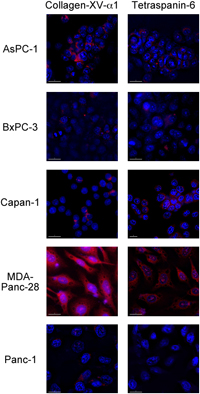
Figure legend:
Immunohistochemical analysis
of five cell lines. Using fluorescently stained antibodies against
collagen-XV-α1 and tetraspanin-6, only cell line MDA-Panc-28 exhibited
strong
signal intensities, which is consistent with the microarray data and
indicates
the specific expression of the two proteins in cells of acinar origin.
The
other four cells are of ductal lineage.
|
FINISHED PROJECT:
Single-step
procedure for the isolation of proteins at near-native conditions from
mammalian tissue for proteomic analysis
|

The
process of extracting
comprehensive proteome representations is a crucial step for many
proteomic
studies. We developed two single-step extraction buffers - Mix 1 and
Mix 2 - for the
isolation of
proteins from mammalian tissues under native conditions in an effective
and
reproducible manner.
..
Protein
extractions were performed from cell lines BxPC-3
and SU.86.86, rat organs (pancreas, liver, heart and lung) and human
pancreatic
cancer tissues. In comparison to several buffer systems that contained
individual
non-ionic
or zwitterionic detergents as well as to commercial extraction
buffers, the two buffer systems were used. Each contains a detergent
cocktail that includes at least one
polymeric
phenylethylene glycol, a long-chain amidosulfobetaine, cholate and a
zwitterionic detergent. Extracts obtained with the various buffer
systens were analysed for protein quantity and
quality. The two detergent cocktails exhibited superior extraction
capacity. Also, they
demonstrated a substantially higher recovery of
membrane and
compartmental proteins as well as much better preservation of protein
functionality. In addition, they did not interfere with subsequent
analysis
steps such
as labelling, a problem that was observed with other buffers. In
Western blot and antibody microarray assays, they
out-performed
the other buffer systems, demonstrating their usefulness for various
types of
proteomic studies.
Alhamdani et al. (2010) J. Prot. Res. 9, 963-971.
|
|
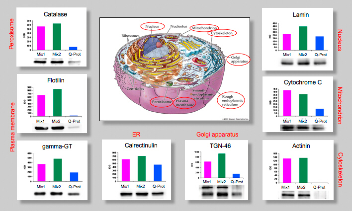
Comparison of protein
extraction efficacy using the two newly established buffer systems and
the best
performing commercial procedure in the analysis. Typical results with
proteins
from different cell compartments are shown.
|
|
|
|
FINISHED PROJECT:
Selection
of highly specific antibodies for the identification of molecular
differences in the proteome of normal and tumour cells |
|
|
|
 |
|
We are
developing systems for
the analysis of complex protein extracts on antibody-microarrays.
Currently, the
availability of specific and highly affine antibodies is a limiting
factor in
such studies. In
collaboration
with the Department
of General Surgery at the University of Heidelberg and Jörg
Hagen of the Proteomics
Unit at Merck in Darmstadt,
we worked at the
establishment of techniques for selecting highly specific antibodies.
..
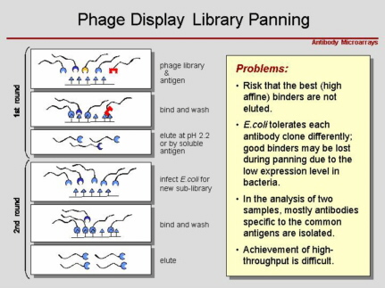
Central
issue to the project was the isolation of highly
specific antibodies from phage
display
libraries. Antibodies isolated from phage display libraries are
typically selected using purified antigens immobilised on plastic
surfaces. In
general,
however, selection requires laborious subtraction protocols to avoid
the selection
of irrelevant antibodies. In spite of such elaborate pre-incubations,
selection
of antibodies by cell panning is limited by background binding of
non-specific phage and relatively low binding of specific phage. Also,
good
binders frequently get lost during the process. Because of the growing
need for
antibodies to be used in early, preventive cancer diagnostics and
tumour specific
therapeutics, we established a process that circumvents the above
mentioned
problems and offers an opportunity selectively to identify highly
specific and
affine antibodies. In addition, since all steps occur in vitro, the technology allows
automatisation of the entire process, which is critical for large-scale
applications.
Hoheisel (2005) PCT / EP2005 / 008431.
|
FINISHED PROJECT:
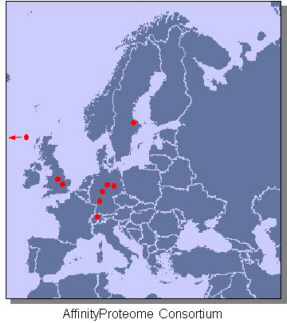
AffinityProteome:
Advanced
affinity tools and technologies
for high-throughput studies of
the human proteome
|
|
High-specificity
affinity reagents (‘binders’) are essential probes for proteome
research, enabling the detection and localisation of multiple proteins
in
tissues and fluids in health and disease through the application of
binder-based technologies (affinity proteomics). This project linked
the
high-throughput production of quality-controlled, recombinant binding
molecules
of different types (antibody fragments, engineered scaffolds, aptamers)
with
advanced applications (capture microarrays, multidimensional
fluorescence
imaging, single-molecule detection, intracellular knockdown) in the
analysis of
human proteome targets.
..
The
partners were five established SMEs and five academic groups engaged in
binder
production and characterisation or protein detection technologies. The
project
aimed at resolving bottlenecks in high-throughput binder production and
application technologies, including cost, throughput, automation and
quality. The
linkage of production, quality control and applications was
exemplified by
targeting proteins involved in signal transduction pathways. These
pathways are
implicated in many diseases. Also, drug development is actively
evolving in this area. Therefore, access to
high-quality
binding
reagents is particularly needed.
..
The
project benefited from close interaction with the FP6 Coordination
Action ProteomeBinders, which
commenced earlier and provided also a framework for activities such as
the ongoing Affinomics project.
|
|
|
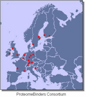
FINISHED PROJECT:
ProteomeBinders: A
European infrastructure of ligand binding molecules against the human
proteome
..
 |
For the
characterisation of the human proteome,
it will
be essential to establish a comprehensive, characterised and
standardised
collection of specific
ligand binding
molecules directed systematically against all the
individual
proteins and
their variant forms. Ligand
binders, which include native and recombinant
antibodies, engineered protein scaffolds, peptides and nucleic acid
aptamers,
are essential reagents for monitoring protein expression and function.
Establishing a binder collection is not an end in itself, but must be
accompanied by development of high-throughput assay systems and look
towards
applications in functional analysis, diagnostics and therapeutics.
..
ProteomeBinders was a
pan-European consortium of 26 European and 2 US partners and had
the remit of networking, database construction and coordination of the
systematic
development, resource management and quality control for these
important
reagents. It combined leading European scientists with complementary
expertise in order to support
systematic
generation and exploitation of binders. Also, the
consortium was integrating
existing infrastructures, reviews technologies, and standardised tools
and
applications.
..
Aiming
ultimately at the
production, characterisation and collection of very many thousands of
specific
and affine binders, the ProteomeBinders resource brought
about benefits for basic and applied research, impacting on healthcare,
diagnostics,
target discovery for drug intervention and therapeutics. For more
detailed
information on the participants, the different techniques and methods
employed
as well as the various acitivties of the consortium, please click on
the logo,
the map or here. |
|
|
|
|
|
Taussig et al. (2007) Nature
Meth. 4, 13-17. |
|
|
|
|
|
Gloriam et al. (2010) Mol. Cell. Prot. 9, 1-10. |
|
|
|
|
|
Alhamdani et al.
(2010) J. Prot. Res. 9, 963-971. |
|
|
|
|
|
Schröder et al. (2010) Antibody Engineering, Vol. 2,
SpringerVerlag, 429-445. |
|
|
|
|
|
Schröder et al. (2010) Mol. Cell. Prot.
9, 1271-1270. |
|
|
|
|
|
Alhamdani et al.
(2010) Proteomics 10, 3203-3207. |
|
|
|
|
|
|
|
|
|
FINISHED PROJECT:
Creation
of an antibody microarrays for the
analysis of the expression of cancer associated proteins |
|

|
|
|
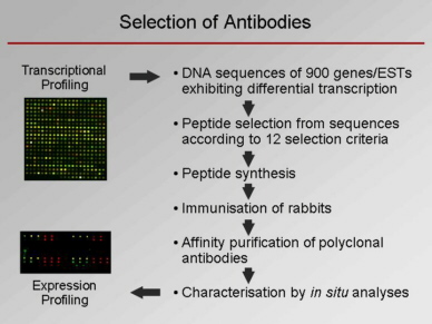
On
the
basis of transcriptional
profiling experiments on DNA-microarrays and other results, some 900
genes of interest were selected. Antibodies for
the
respective
proteins were generated in a collaboration with the company Eurogentec by
selecting and
synthesising appropriate peptide sequences.
The peptides were used for the immunisation of rabbits, from which
affinity-purified antibodies were obtained.
..
Further
characterisations
were performed to define specificity and affinity of the
molecules.This process provided us
with an initial set
of
some 650 antibodies
that have been used in a large number of experiments, validating their
value and performance. We continue using them as probes on microarrays
and beyond for analyses of protein
expression variations. Further
antibodies with particular
characteristics are being produced and checked for their suitability
for
cancer diagnosis.
..
With
the existing
microarray, we pursue biomedical studies toward an early and accurate
diagnosis on tissues, cells and various forms of body fluids. In
addition, we are aiming at understanding protein-based communication
between the cells of the tumour microenvironment. Analyses are under
way
comparing transcriptional changes and the actual variations at the
protein
level. Also other applications are being worked at.
..
Apart
from the
production of antibodies by classical means, other procedures for the
isolation
of antibodies are being pursued. Activities in the group are mainly
based
on
display libraries. In addition, we collaborate
with
partners within the Affinomics consortium, who
actively produce binders of different formats.
Schröder et al. (2010) Mol. Cell. Prot.
9, 1271-1270.
|
|
|
FINISHED PROJECT:
Subcellular
protein extraction from human pancreatic cancer tissue
Proteins are
the major class
of effector molecules in cellular systems. For the identification of
functional
differences between normal and diseased tissues, a reliable analysis of
their
protein content is essential. Reproducible isolation and fractionation
of
intact proteins are important in this respect. The complexity of
proteins in
structure and concentration, their close interaction as well as their
instability represent major challenges. For isolation from tissues,
also the
destruction of the cell-cell and cell-matrix connections within a
tissue
without affecting protein quality is a critical factor. We compared
different
processes for a compartmental protein preparation from pancreatic
tissues, using matching normal and tumour tissue samples from the same
patients.
Pancreas is one of the most challenging tissues because of its high
content of
proteases.
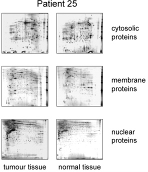
..
Success of the different procedures varied strongly.
Based on a
scheme of slicing the tissues and a subsequent isolation of the cells,
we
established a workflow for the extraction in a reproducible manner of
fractions
of cytosolic proteins, membrane and organelle proteins, nuclear
proteins and
cytoskelatal filaments. The tissue slices also allow for a
representative
confirmation of the individual samples’ cellular status by
histochemical
processes and a proper separation or mixing of cellular material from
across a
tumour if required.
Börner et al. (2009) BioTechniques 46, 297-304.
|
|
 |
|
FINISHED PROJECT:
Appropriate
design of microspot immunoassays;
compensation for kinetic limitations
We
examined the limitations of existing microarray immunoassays and
investigated
how best to optimise them using theoretical and experimental
approaches. A key
physicochemical limitation of microarray immunoassays is a strong
dependence of
antibody microspot kinetics on the mass flux to the spots. We analysed
theoretically and experimentally the effects of microarray design
parameters
(incubation vessel geometry, incubation time, stirring, spot size,
antibody-binding site density, etc.) on microspot reaction kinetics and
sensitivity.
..
Using a two-compartment model, the quantitative
descriptors of the
microspot reaction were determined for different incubation and
microarray
design conditions. This analysis revealed profound mass transport
limitations
in the observed kinetics, which may be slowed down as much as hundreds
of times
compared with solution kinetics.
..
The data obtained were considered with
relevance to microspot assay diffusional and adsorptive processes,
enabling us
to validate some of the underlying principles of the antibody microspot
reaction mechanism and provide guidelines for optimal microspot
immunoassay
design. For assays optimised to maximize the reaction velocity, we
could
demonstrate sensitivities in the attomolar and low femtomolar ranges.
|
|
|
|
|
|
|
|
|
Kusnezow et al.
(2005) Handbook of Immunohistochemistry, Vol. 2 (Hayat, M.A.,
ed.),
Elsevier, 23-35. |
|
|
|
|
Klenin et
al. (2005) J.
Chem. Phys. 122, Art. No. 214715. |
|
|
|
|
Syagailo
et al. (2006) Apoptosis
and Cancer Therapy (Debatin, K.M. & Fulda, S., eds.),
Wiley-VHC, 60-85. |
|
|
|
|
Kusnezow et
al. (2006) Proteomics 6, 794-803. |
|
|
|
|
Kusnezow et
al. (2006) Expert
Rev. Mol. Diagn. 6, 111-124. |
|
|
|
|
Kusnezow et
al.
(2006) Mol. Cell. Prot. 5, 1681-1696. |
|
|
|
|
|
|
FINISHED PROJECT:
Antibody
microarrays: production parameters |

|
Antibody
microarrays will have
an enormous impact on the functional analysis of cellular activity and
regulation, especially at the level of protein expression and
protein-protein
interaction. The array surface is bound to have a tremendous influence
on the
findings from such studies.
..
Apart from
the basic issue of
how to attach antibodies optimally without affecting their function, it
is also
important that the cognate antigens, applied within a complex protein
mixture,
all bind to the arrayed antibodies irrespective of their enormous
variety in
structure.
..
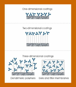
We analysed
various factors in
the production of antibody microarrays on glass support – the
modification of
the glass surface, kind and length of crosslinkers, composition and pH
of the
spotting buffer, blocking reagents, antibody concentration, storage
procedures,
etc. – in order to evaluate their effect on array performance.
Altogether, data
from more than 1200 individual array experiments was taken into
account. In
addition to home-made slides, also commercially available systems were
included
in the analysis. The results of this work can be found in the
manuscripts
listed below.
..
Further
improvements were made
more recently and are documented on the current webpage.
..
|
|
|
Kusnezow & Hoheisel
(2002) BioTechniques 33 (suppl.), 14-23. |
|
|
|
|
Kusnezow et
al. (2003) Proteomics 3, 254-264. |
|
|
|
|
Kusnezow & Hoheisel
(2003) J. Mol. Recognit. 16,
165-176. |
|
|
|
|
Bauer et al.
(2003) Comp. Funct. Genome 4,
520-524. |
|
|
|
|
Kusnezow et al.
(2004) Protein Microarrays (Schena, M., ed.), Jones and
Bartlett,
247-284. |
|
|
|
|
|
|
|
|
|