Breast Cancer Awareness Month October: Will imaging replace biopsies in cases of suspected breast cancer?
Each year, approximately 2.8 million women have a mammography as part of the breast cancer screening program in Germany. In about 35,000 of these cases, the procedure reveals a suspicious finding that physicians follow up on by taking a tissue sample. Only half of these women, however, truly have breast cancer. Scientists from the German Cancer Research Center (DKFZ), in collaboration with mammography screening units in Heidelberg and Mannheim, have now published the first data to suggest that in many cases, advanced diffusion-weighted MR imaging can help women avoid an unnecessary control biopsy.
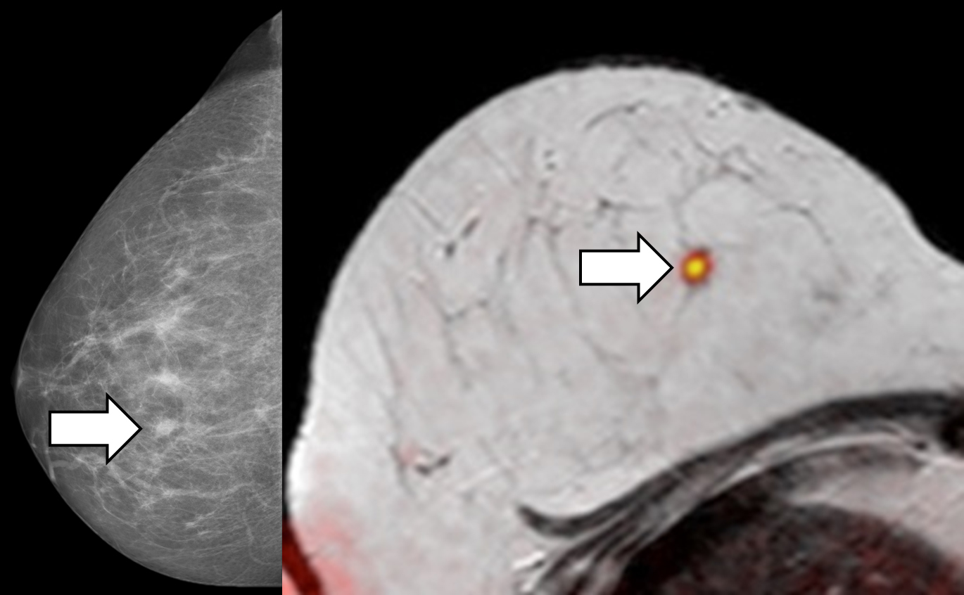
Suspicious lesion detected in the X-ray screening mammogram (left image). The lesion is confirmed using the combined diffusion weighted breast MR technique with the orange signal suggesting possible malignancy. Histopathology confirmed a malignant breast tumor.
© dkfz.de
Statistically, about one in 20 women who undergo mammography screening can expect a suspicious finding. If further tests indicate a possibility of cancer, the screening physician recommends taking a tissue sample, or biopsy. Nearly 35,000 women every year face this situation. However, in only about 17,000 of these cases is a malignant tumor actually found, says Dr. Sebastian Bickelhaupt, a radiologist at the DKFZ who has been investigating the use of advanced MR imaging in diagnosing breast cancer. We have been looking at advanced imaging technologies as a potential way of reducing the number of invasive tissue examinations.
In a mammogram, which examines the breast using X-rays, it is often impossible to distinguish benign from malignant abnormalities in tissues and thus exclude the existence of a malignant tumor to the physician's satisfaction. If the situation can't be clarified by further testing, such as an ultrasound examination, an invasive biopsy must be performed.
The DKFZ radiologists have optimized a method called diffusion-weighted magnetic resonance imaging (MRI) specifically for use in these cases. Diffusion-weighted MRI is a special technique that allows us to see the movement of water molecules in tissues, explains Professor Heinz-Peter Schlemmer, head of Radiology at the DKFZ. Since tumors strongly reduce the movement of these molecules, we decided to examine the potential of our optimized breast MRI for deeper investigations of suspicious findings, in hopes of avoiding an unnecessary biopsy.
This idea led the DKFZ researchers to plan a study in close collaboration with the office-based physicians in Dr. Wolfgang Lederers team at Heidelberg ATOS Klinik and Dr. Heidi Daniels team at the Radiology Center Mannheim, who routinely conduct mammography screenings. If a mammography shows a suspicious lesion, the patient is invited to the Radiology Center Mannheim for further testing and, as a rule, also for biopsies.
For our study, we asked affected women if they were prepared to have an optimized breast MRI prior to the biopsy, Daniel explained. We were surprised to get such a high rate of participation that we could proceed with the study quickly," Lederer adds. We owe our thanks to the many participants, Lederer adds.
The DKFZ radiologists compared the MRI images with results from the biopsies. Within the first 50 cases we investigated, we were already thrilled, says Bickelhaupt. Adding the step of optimized breast MRI enabled us to classify over 90 percent of the suspicious findings correctly. That's an enormous improvement over the 50-percent rate achieved by a combination of mammography and a subsequent ultrasound examination.
In Schlemmers opinion, this does not mean that breast MRI is ready to replace screening mammography. The positive results of our study are based on using MRI in combination with other tests. X-ray mammography also detects minute microcalcifications that indicate non-invasive breast cancer (DCIS), which do not appear in MRI. According to Schlemmer, optimized breast MRI is most suitable for clarifying a suspicious finding. A biopsy would only be required if the MRI indicates a high likelihood for a positive finding.
The scientists enhanced and optimized diffusion-weighted MRI specifically for their study. In collaboration with colleagues from the German Cancer Research Center, they established a quality management system to standardize and ensure the quality of breast MRI that can be used on all standard MR devices.
We are very grateful to the Dietmar Hopp Foundation, whose generous support made our study possible, added Schlemmer. If the results are confirmed over the further course of our study, we are well on our way to reducing the enormous emotional stress experienced by women whose screening mammograms yield unclear findings.
The scientists have now published their promising intermediate results in the US journal Radiology. We expect to be able to examine the 250 cases foreseen in the study by October, and we hope, of course, that our initial results will be confirmed, says Bickelhaupt. Substantially larger studies will have to follow before physicians can refrain from performing biopsies on the basis of MRI scans alone, and before health insurance plans assume the costs of diffusion-weighted MRI.
The DKFZ radiologists now plan to investigate whether diffusion-weighted MRI can also be used in other types of cancer to clarify suspicious findings and monitor the progress of therapies.
Bickelhaupt, S; Laun, F; Tessdorf, J; Lederer, M; Daniel, H; Stieber, A; Delorme, S; Schlemmer, HP: Fast and Noninvasive Characterization of Suspicious Lesions Detected at Breast Cancer X-Ray Screening: Capability of Diffusion-weighted MR Imaging with MIPs1. Radiology 2015, DOI 10.1148/radiol.2015150425
Pictures for this press release are available on the Internet at:
http://www.dkfz.de/de/presse/pressemitteilungen/2015/bilder/boesartig.jpg
Caption: Suspicious lesion detected in the X-ray screening mammogram (left image). The lesion is confirmed using the combined diffusion weighted breast MR technique with the orange signal suggesting possible malignancy. Histopathology confirmed a malignant breast tumor. Source: DKFZ
http://www.dkfz.de/de/presse/pressemitteilungen/2015/bilder/Entwarnung.jpg
Caption: A suspicious lesion that was detected in the X-ray screening mammogram (not displayed) is displayed in conventional MR-Mammography (upper image). Additive diffusion weighted imaging (DWI) sequences suggests the lesion to potentially be benign (lower image). Biopsy revealed the lesion to be benign. Source: DKFZ
With more than 3,000 employees, the German Cancer Research Center (Deutsches Krebsforschungszentrum, DKFZ) is Germanys largest biomedical research institute. DKFZ scientists identify cancer risk factors, investigate how cancer progresses and develop new cancer prevention strategies. They are also developing new methods to diagnose tumors more precisely and treat cancer patients more successfully. The DKFZ's Cancer Information Service (KID) provides patients, interested citizens and experts with individual answers to questions relating to cancer.
To transfer promising approaches from cancer research to the clinic and thus improve the prognosis of cancer patients, the DKFZ cooperates with excellent research institutions and university hospitals throughout Germany:
The DKFZ is 90 percent financed by the Federal Ministry of Education and Research and 10 percent by the state of Baden-Württemberg. The DKFZ is a member of the Helmholtz Association of German Research Centers.