HP-F4: Biomedical Imaging and Radiation Oncology
Type: Practical Course with Student Seminars
Date: 8.-26. January 2024
Location: REZ (Radiologisches Forschungs- und Entwicklungszentrum DKFZ), INF 223; 1st floor seminar room and laboratories
Hosts/Supervisors: Mark E. Ladd, Ina Kurth and colleagues from Research Program E 'Imaging and Radiooncology'
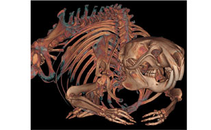
In vivo Bildgebung einer Ratte mittels Flächendetektor Volumen Computertomographie (flat panel volumetric computed tomography, Volume CT Siemens). Dreidimensionale Rekonstruktion eines Rattenskeletts in hoher räumlicher Auflösung
© dkfz.de
Topics:
Non-invasive imaging is crucial for the diagnosis and therapy monitoring of cancer and plays an increasing role in preclinical research. In addition, image guided radiotherapy concepts have broadly been established in the clinics. The aim of this course is to introduce the most relevant imaging and radiotherapy strategies as well as radiopharmaceutical and molecular radiobiological principles used in oncology.
Content:
After the course the students will be familiar with the basic principles of computed tomography, magnetic resonance imaging, ultrasound, optical imaging, PET and SPECT as well as the application of most frequently used contrast agents in clinical settings and in preclinical research. In this context, it will also be explained how these imaging modalities can be used for molecular and cellular imaging. Additionally, basic principles of radiotherapy planning, treatment and therapy monitoring including the concept of precision therapy will be explained.